

Succinate dehydrogenase gene mutations in cardiac paragangliomas
- PMID: 25896150
- PMCID: PMC4450109
- DOI: 10.1016/j.amjcard.2015.03.020
Succinate dehydrogenase gene mutations in cardiac paragangliomas
Abstract
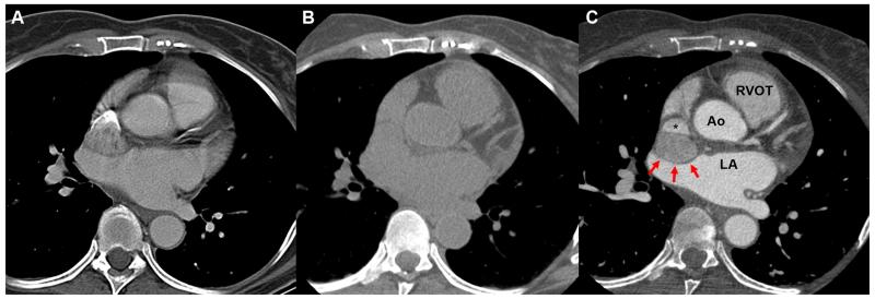
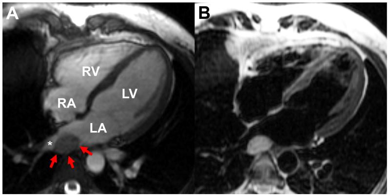
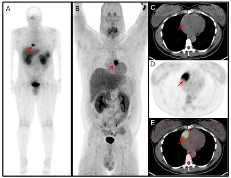
Pheochromocytomas and paragangliomas are chromaffin cell tumors arising from neuroendocrine cells. At least 1/3 of paragangliomas are related to germline mutations in 1 of 17 genes. Although these tumors can occur throughout the body, cardiac paragangliomas are very rare, accounting for <0.3% of mediastinal tumors. The purpose of this study was to determine the clinical characteristics of patients with cardiac paragangliomas, particularly focusing on their genetic backgrounds. A retrospective chart analysis of 15 patients with cardiac paragangliomas was performed to determine clinical presentation, genetic background, diagnostic workup, and outcomes. The average age at diagnosis was 41.9 years. Typical symptoms of paraganglioma (e.g., hypertension, sweating, palpitations, headache) were reported at initial presentation in 13 patients (86.7%); the remaining 2, as well as 4 symptomatic patients, initially presented with cardiac-specific symptoms (e.g., chest pain, dyspnea). Genetic testing was done in 13 patients (86.7%); 10 (76.9%) were positive for mutations in succinate dehydrogenase (SDHx) subunits B, C, or D. Thirteen patients (86.7%) underwent surgery to remove the paraganglioma with no intraoperative morbidity or mortality; 1 additional patient underwent surgical resection but experienced intraoperative complications after removal of the tumor due to co-morbidities and did not survive. SDHx mutations are known to be associated with mediastinal locations and malignant behavior of paragangliomas. In this report, the investigators extend the locations of predominantly SDHx-related paragangliomas to cardiac tumors. In conclusion, cardiac paragangliomas are frequently associated with underlying SDHx germline mutations, suggesting a need for genetic testing of all patients with this rare tumor.
Published by Elsevier Inc.
Figures
References
-
- Lenders JWM, Eisenhofer G, Mannelli M, Pacak K. Phaeochromocytoma. Lancet. 2005;366:665–675. - PubMed
-
- Eisenhofer G, Lenders JWM, Timmers H, Mannelli M, Grebe SK, Hofbauer LC, Bornstein SR, Tiebel O, Adams K, Bratslavsky G, Linehan WM, Pacak K. Measurements of plasma methoxytyramine, normetanephrine, and metanephrine as discriminators of different hereditary forms of pheochromocytoma. Clin Chem. 2011;57:411–420. - PMC - PubMed
-
- Gimenez-Roqueplo AP, Dahia PL, Robledo M. An update on the genetics of paraganglioma, pheochromocytoma, and associated hereditary syndromes. Horm Metab Res. 2012;44:328–333. - PubMed
-
- Whalen RK, Althausen AF, Daniels GH. Extra-adrenal pheochromocytoma. J Urol. 1992;147:1–10. - PubMed
-
- Patel J, Sheppard MN. Pathological study of primary cardiac and pericardial tumours in a specialist UK Centre: surgical and autopsy series. Cardiovasc Pathol. 2010;19:343–352. - PubMed
Publication types
MeSH terms
Substances
Grants and funding
LinkOut - more resources
Full Text Sources
Other Literature Sources
