. 2014;58(9):693-9.
doi: 10.1387/ijdb.140288jp.

Construction and characterization of a sox9b transgenic reporter line

Affiliations

Construction and characterization of a sox9b transgenic reporter line

Jessica S Plavicki et al. Int J Dev Biol. 2014.

Abstract

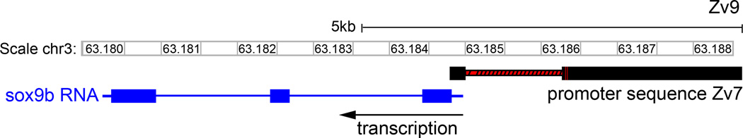
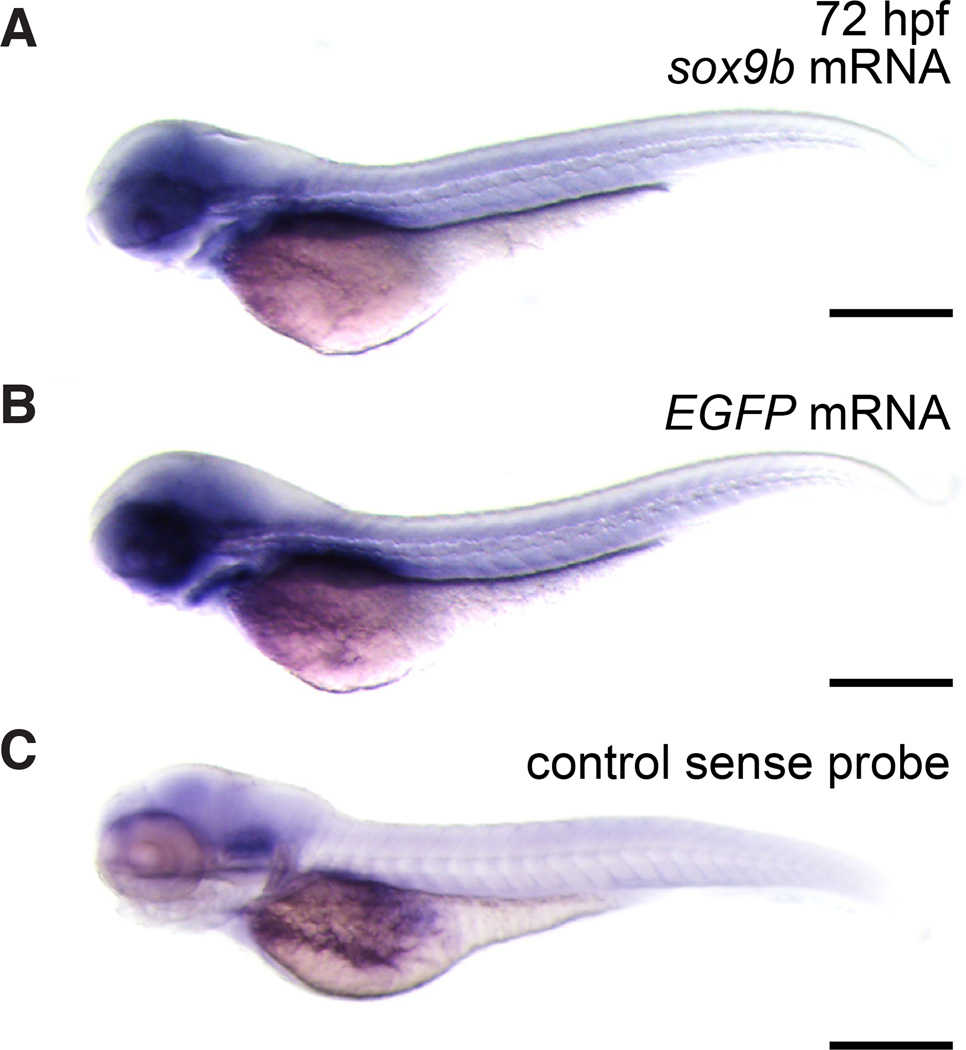
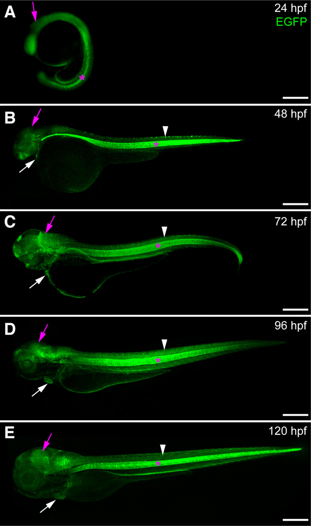
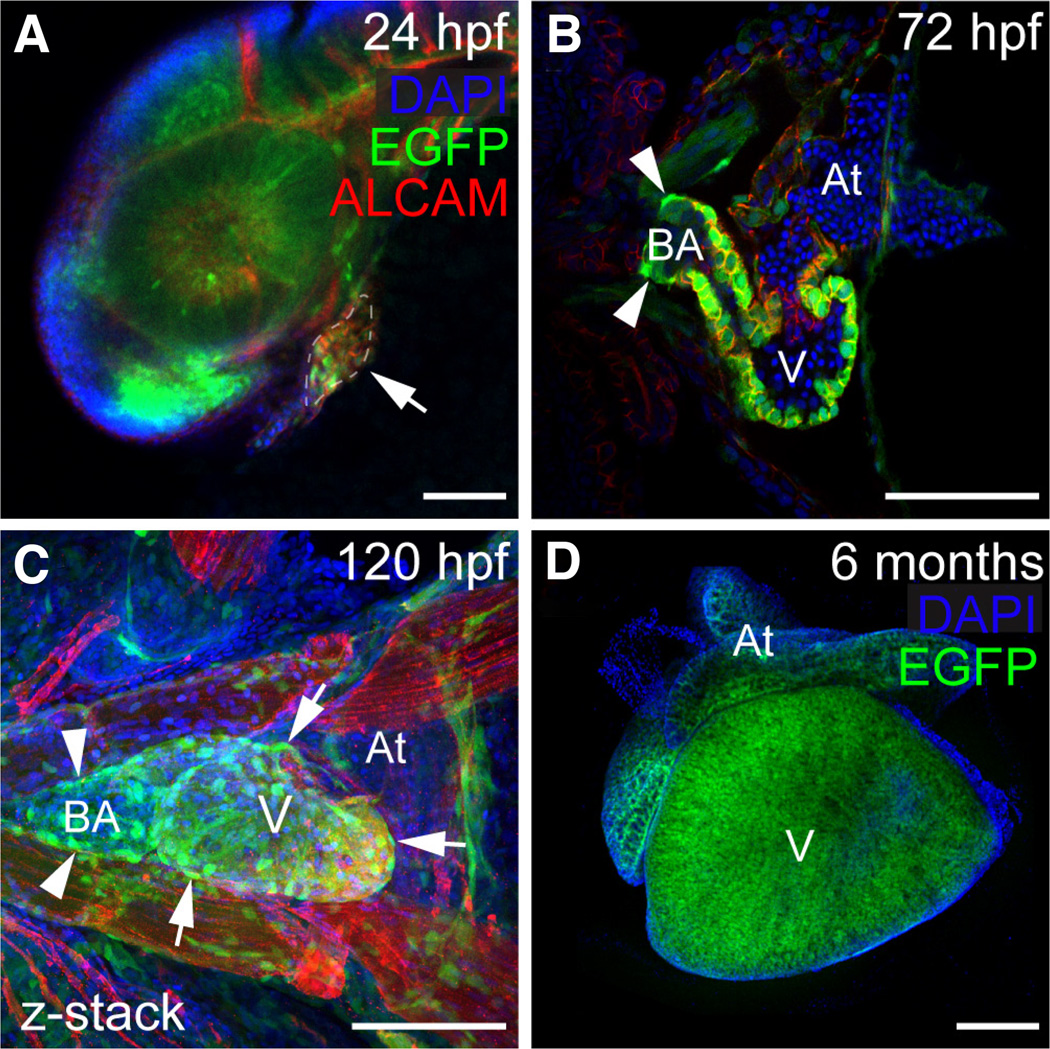
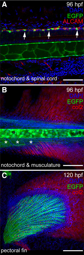
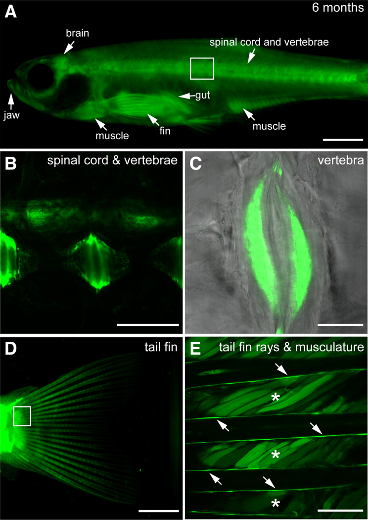
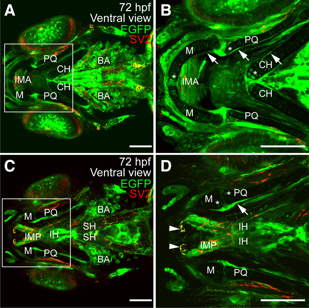
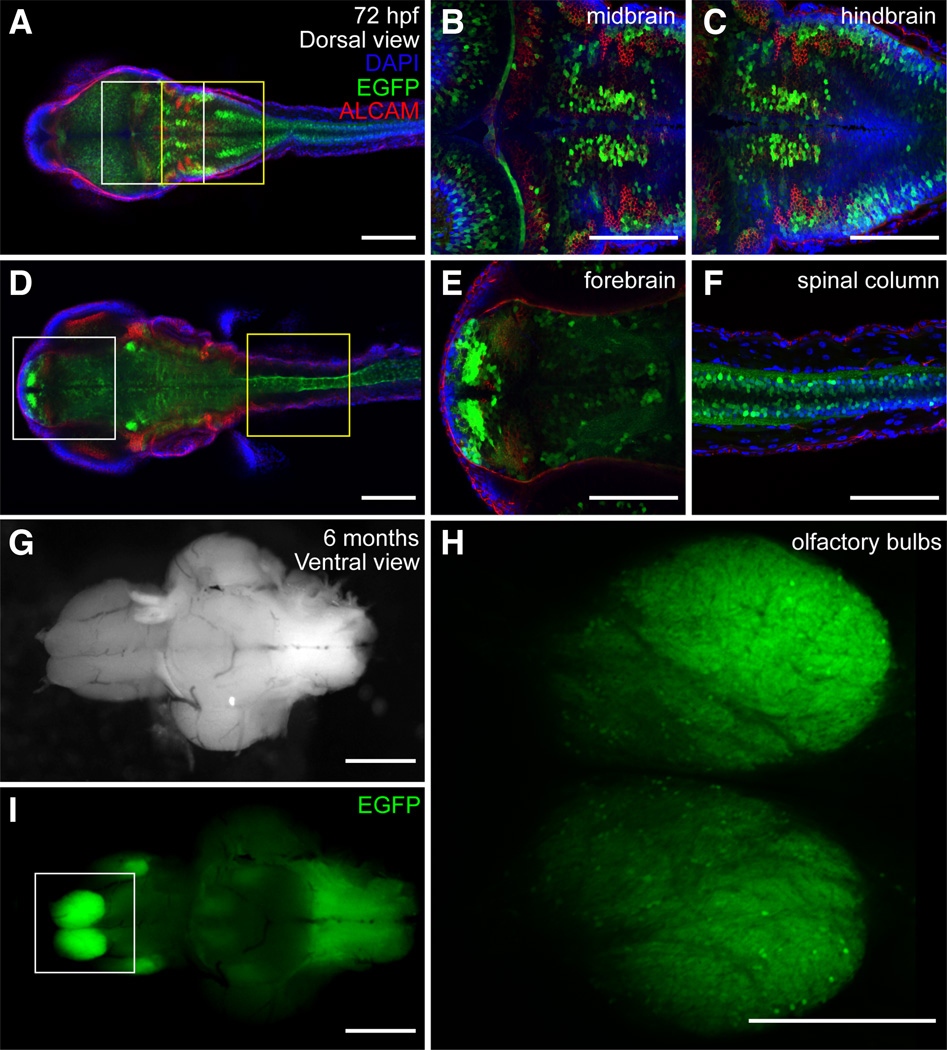
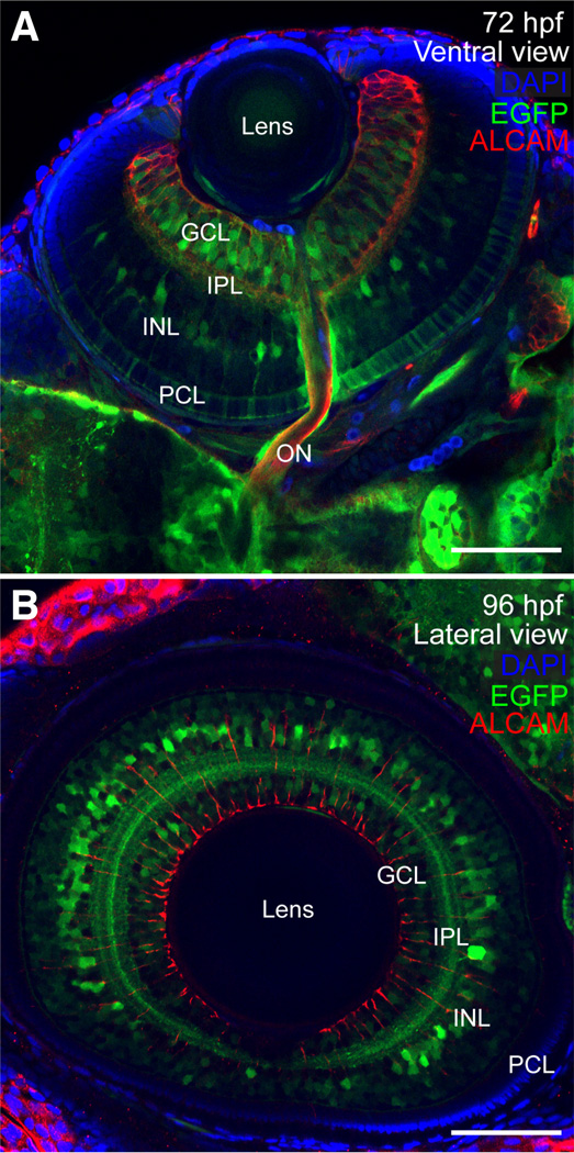
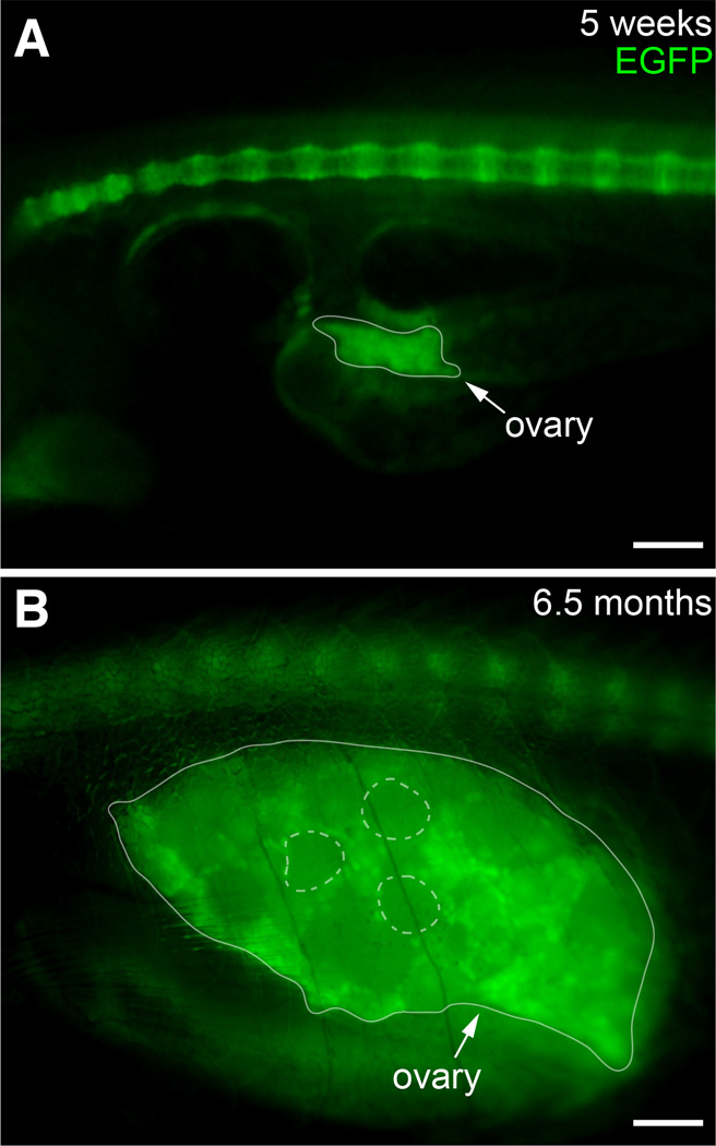
The transcription factor SOX9 is a member of the SRY-related high-mobility-group box (SOX) superfamily of genes. In mammals, Sox9 plays important roles in many developmental processes including craniofacial, skeletal and heart morphogenesis, retinal and brain development, and gonad differentiation. Human mutations in SOX9 or the SOX9 promoter result in campomelic dysplasia, a severe genetic disorder, which disrupts skeletal, craniofacial, cardiac, neural and reproductive development. Due to the duplication of the teleost fish genome, zebrafish (Danio rerio) have two Sox9 genes: sox9a and sox9b. Loss of sox9b in zebrafish results in loss of function phenotypes that are similar to those observed in humans and mice. In order to generate a transgenic sox9b:EGFP reporter line, we cloned a 2450 bp fragment of the sox9b promoter and fused it to an EGFP reporter. Consistent with reported sox9b expression and function, we observed sox9b:EGFP in the developing heart, skeletal and craniofacial structures, brain, retina, and ovaries. Our resulting transgenic line is a useful tool for identifying and studying sox9b function in development and visualizing a number of zebrafish organs and tissues in which sox9b is normally expressed.


Figures

Fig. 1
Fig. 1. Schematic comparing the cloned 2450 sox9b promoter sequence in builds Zv7 and Zv9
BLAT of the cloned 2450 bp sox9b promoter sequence in build Zv7 with the sequence in build Zv9. Both a short region adjacent to the transcriptional start site and a ~ 1.1 kb region align well. However, approximately 1.4 kb has no alignment in build Zv9 and has been replaced by a unique sequence. Solid black bars in the Zv7 promoter sequence schematic indicate areas of sequence alignment. Red regions indicate areas where the sequences in Zv7 and Zv9 do not align.
Fig. 2
Fig. 2. sox9b transgenic line recapitulates the endogenous pattern of sox9b expression
(A–C) Lateral views of 72 hpf embryos. To detect sox9b, in situ hybridization was performed on whole embryos. (A) sox9b mRNA in transgenic embryos. (B) EGFP mRNA in transgenic embryos. (C) A sense probe was used as a control for nonspecific binding. Scale bars, 100 microns.
Fig. 3
Fig. 3. Expression of sox9b:EGFP during embryonic and larval development
(A–E) Lateral views of sox9b:EGFP embryos and larvae. (A) Epifluorescent image at 24 hpf. (B–E) Confocal images at 48 hpf (B), 72 hpf (C), 96 hpf (D) and 120 hpf (E). sox9b:EGFP expression is detected in the brain (purple arrows), eye, heart (white arrows), jaw, spinal cord (white arrowhead) and notocord (pink asterisks). Scale bars, 100 microns.
Fig. 4
Fig. 4. Expression of sox9b:EGFP in the heart
(A–D) Confocal images of sox9b:EGFP expression in embryonic, larval, and adult zebrafish hearts. Cardiomyocytes are marked with activated leukocyte cell adhesion molecule (ALCAM; red in A–C) and nuclei are stained with DAPI (blue); (A) Lateral view of a 24 hpf embryo. The developing heart is outlined and indicated by the arrow. (B,C) Ventral views of a 72 and 96 hpf hearts. Expression of sox9b:EGFP is detected in cardiomyocytes, the bulbus arteriosus (arrowheads in B and C), and in epicardial cells (arrows in C). (D) Ventral view of an adult heart. Expression of sox9b:EGFP in cardiomyocytes continues into adulthood. V is ventricle; At, atrium; BA, bulbus arteriosus. Anterior is to the left in all panels. Scale bars, 50 microns.
Fig. 5
Fig. 5. Expression of sox9b:EGFP in larval spine, skeleton, and fin
(A–C) Lateral views of a 96 hpf and 120 larvae. Confocal images of sox9b:EGFP expression (green) in zebrafish spinal cord (A), notochord (A,B), and pectoral fin (C). (A) Neurons in the spinal cord are marked with activated leukocyte cell adhesion molecule (ALCAM; red in A). Expression of sox9b:EGFP is seen in a subset of neurons (arrows) and in the notochord. (B) sox9b:EGFP is also expressed in muscle associated with the spine (asterisks in B) and the developing pectoral fin. Collagen 2 (col2; red in B,C) is expressed in the fins. (A–C) Nuclei are stained with DAPI (blue). Scale bars, 50 microns.
Fig. 6
Fig. 6. Expression of sox9b:EGFP in adult skeleton and skeletal muscle
(A–E) Lateral views of six month old sox9b:EGFP transgenic fish. (B) Area boxed in (A). Expression is detected in the spinal cord and vertebrae. (C) Magnification of the sox9b:EGFP expressing growth plates in the vertebrae. (D) Muscle associated with the tail fin and rays within the tail fin express sox9b:EGFP. (E) Magnification of the box in (D). Asterisks mark the muscle while arrows point to the rays. Scale bars in (A,D), 150 microns. Scale bars in (B,C,E), 50 microns.
Fig. 7
Fig. 7. Expression of sox9b:EGFP in larval jaw and associated skeletal muscle
(A–D) Ventral views of 72 hpf larvae stained for synaptic vesicles (SV2; red). sox9b:EGFP expression was detected in the perichondrium and in chondrocytes. (B,D) Magnification of regions in the white boxes in panels (A,C) respectively. Expression is associated with the ceratohyal (CH), Meckel’s (M), palatoquadrate (PQ), ceratobranchials (not shown) and basihyal (not shown) cartilages. Expression was also detected in muscles associated with the jaw including the intermandibularis anterior (IMA), intermandibularis posterior (IMP), interhyoideus (IH), and sternohyoideus (SH). sox9b:EGFP was also observed in the branchial arches (BA). Scale bars, 50 microns.
Fig. 8
Fig. 8. Expression of sox9b:EGFP in larval and adult brain
(A–F) Confocal images showing dorsal views of a 72 hpf larval zebrafish. Larvae were stained for ALCAM (red) and mounted in Vectashield with DAPI (blue). (B,C) Magnification of the regions in the white and yellow boxes in (A). (E,F) Magnification of the regions in the white and yellow boxes in (D). Expression of sox9b:EGFP in the fore-, mid- and hindbrain as well as in the spinal cord. (G,I) Bright field images and epifluorescence and ventral views of an adult brain. (H) Magnification of olfactory bulb boxed in (I). Scale bars, 50 microns.
Fig. 9
Fig. 9. Expression of sox9b:EGFP in retina
(A,B) Ventral and lateral views of larval eyes at 72 and 96 hpf stained for ALCAM (red) and mounted in Vectashield with DAPI (blue). (A) At 72 hpf, sox9b:EGFP expression was observed in a subset of cells in the photoreceptor cell layer (PCL), inner nuclear layer (INL), and ganglion cell layer (GCL) and within the inner plexiform layer (IPL). (B) By 96 hpf, sox9b:EGFP expression in the PCL was waning, however sox9b:EGFP was visible in the INL, IPL, and GCL. Scale bars, 50 microns.
Fig. 10
Fig. 10. Expression of sox9b:EGFP in ovary
Lateral views of juvenile (A) and adult (B) ovary. The ovaries are outlined with a solid line. Examples of mature follicles are outlined with dashed circles. Scale bars, 100 microns.

References

    1. Chiang EFL, Pai CI, Wyatt M, Yan YL, Postlethwait J, Chung BC. Two sox9 genes on duplicated zebrafish chromosomes: Expression of similar transcription activators in distinct sites. Dev Bio. 2001;231:149–163. - PubMed
    1. Esain V, Postlethwait JH, Charnay P, Ghislain J. FGF-receptor signalling controls neural cell diversity in the zebrafish hindbrain by regulating olig2 and sox9. Development. 2010;137:33–42. - PMC - PubMed
    1. Foster JW, Dominguez-Steglich MA, Guioli S, Kwok C, Weller PA, Stevanovic M, Weissenbach J, Mansour S, Young ID, Goodfellow PN, Brook JD, Schafer AJ. Campomelic dysplasia and autosomal sex reversal caused by mutations in an SRY-related gene. Nature. 1994;372:525–530. - PubMed
    1. Hofsteen P, Plavicki J, Johnson SD, Peterson RE, Heideman W. Sox9b is required for epicardium formation and plays a role in TCDD-induced heart malformation in zebrafish. Mol Pharmacol. 2013;84:353–360. - PMC - PubMed
    1. Houston CS, Opitz JM, Spranger JW, Macpherson RI, Reed MH, GilBert EF, Herrmann J, Schinzel A. The campomelic syndrome: review, report of 17 cases, and follow-up on the currently 17-year-old boy first reported by Maroteaux et al., in 1971. Am J Med Genet. 1983;15:3–28. - PubMed

Publication types

MeSH terms

Substances