Review
. 2015 May;17(5):392-404.
doi: 10.1177/1098612X14568170.

Feline gastrointestinal eosinophilic sclerosing fibroplasia: 13 cases and review of an emerging clinical entity

Affiliations
Review

Feline gastrointestinal eosinophilic sclerosing fibroplasia: 13 cases and review of an emerging clinical entity

Michael Linton et al. J Feline Med Surg. 2015 May.

Abstract

Objective: Feline gastrointestinal eosinophilic sclerosing fibroplasia (FGESF) is a recently described inflammatory disease of cats affecting stomach or intestines and draining regional lymph nodes. This study presents clinical and laboratory data on 13 newly described cases from Australia (11) and the UK (two).

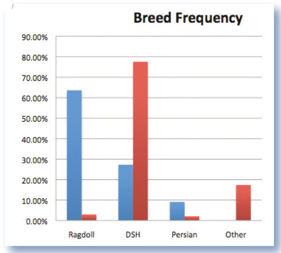
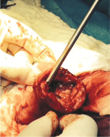
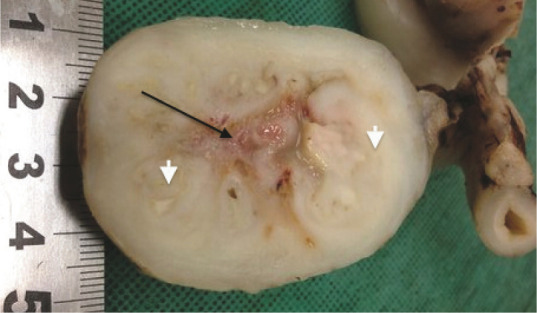
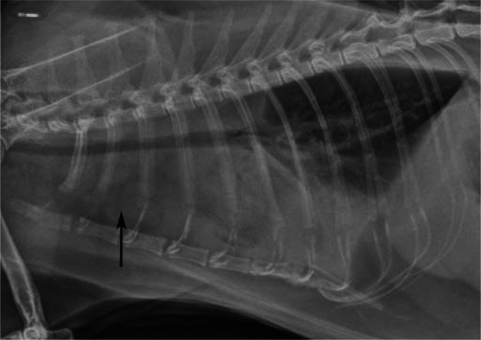
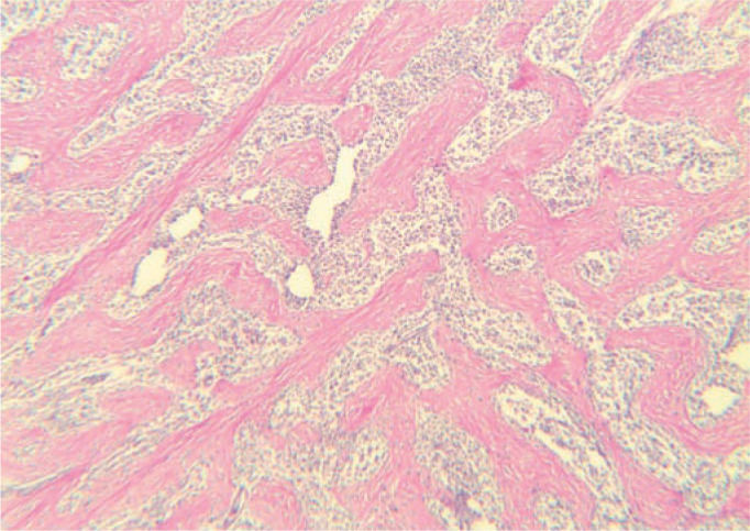
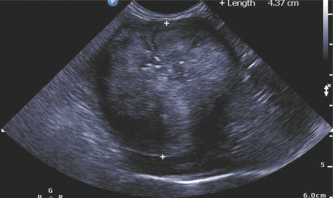
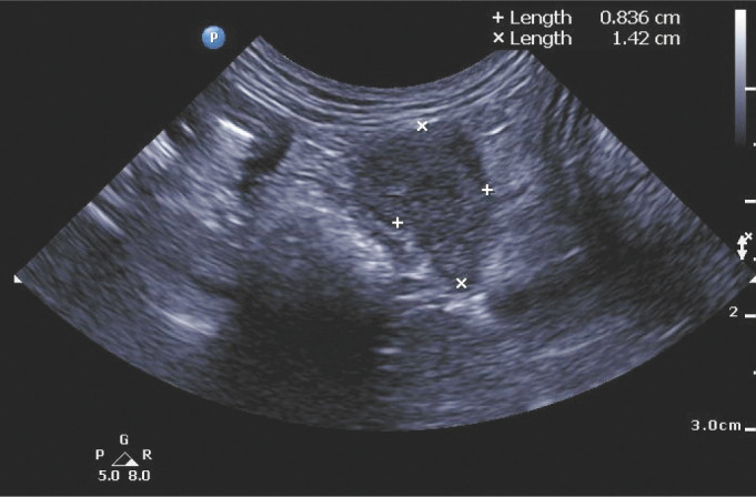
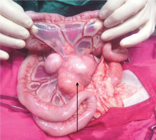
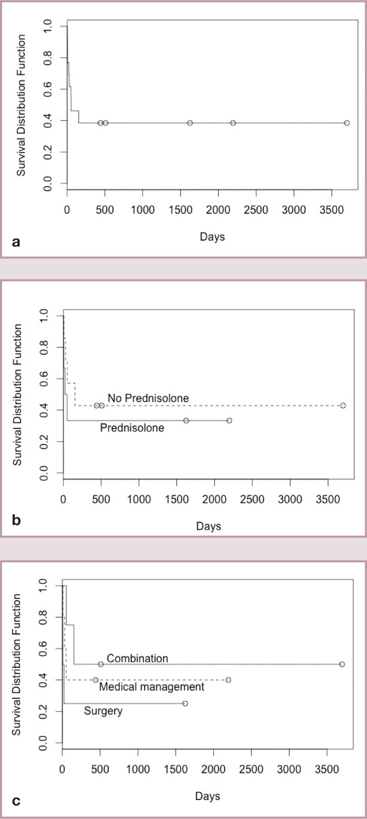
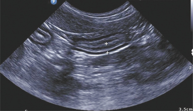
Observations: The disease was most often observed in middle-aged cats (median 7 years of age; interquartile range 5-9 years). Ragdolls (7/13) and males (9/13) were overrepresented. Cats generally had a long history of vomiting and/or diarrhoea. Lesions were typically large, hard, non-painful, easily palpable and most commonly situated near the pylorus or ileocaecocolic junction. Lesions were heterogeneous ultrasonographically and on sectioning at celiotomy or necropsy. Masses were hard and 'gritty' on fine-needle aspiration due to internal trabeculae made up of mature collagen bundles. Bacteria were commonly detected within masses (9/13 cases) using either culture or conventional light microscopy and a panel of special stains, and/or fluorescence in situ hybridisation (FISH), although detection often required a diligent search of multiple tissue sections. A consistent bacterial morphology could not be appreciated among the different cases.

Outcome: Patients were treated with a variable combination of cytoreduction (debulking and biopsy, to complete surgical resection), immunosuppressive therapy and antimicrobial agents. Many cats had a poor outcome, which was attributable to late diagnosis combined with suboptimal management. It is hoped that suggestions outlined in the discussion may improve clinical outcomes and long-term survival in future cases.


Conflict of interest statement

The authors do not have any potential conflicts of interest to declare.

Figures

Figure 1
Figure 1
Observed frequency of each breed for cats with confirmed FGESF (blue bars). Red bars show expected breed frequencies based on registration data from the Companion Animals Register, New South Wales, Australia. DSH = domestic shorthair
Figure 2
Figure 2
Intraoperative photograph of the pyloric lesion in case 11. Marked thickening of the pyloric wall is evident
Figure 3
Figure 3
Photograph of the resected jejunal mass from case 10. The intestinal lumen has been completely obliterated, resulting in signs of intestinal obstruction. The arrow denotes a region of caseous necrosis; arrowheads identify foci of mature collagen
Figure 4
Figure 4
Lateral thoracic radiograph from case 2 presented for respiratory distress. Note the pleural effusion (most evident cranioventrally) and prominent sternal lymph node (arrow). Although this is an atypical presentation, it is important to emphasise that some cases of FGESF can present with bicavitary involvement, the disease process starting in the abdomen and spreading to the thoracic cavity, presumably due to drainage of abdominal lymphatics to the sternal lymph node. A similar observation was reported recently3
Figure 5
Figure 5
Histology of the resected lesion from case 10. The characteristic network of coarse collagen trabeculae (staining pink) throughout the lesion is an important distinguishing feature of FGESF. This abundance of collagen causes the hard, gritty texture of these lesions, which is most obvious during biopsy procedures (or during dissection at necropsy). Haematoxylin and eosin, × 400 magnification
Figure 6
Figure 6
Ultrasound image of the pyloric lesion (denoted by +) of case 11. Note the loss of layering with the mixture of hypoechoic and hyperechoic regions within the tissue. It is thought that the hyperechoic regions correspond with the fibrotic zones described histologically
Figure 7
Figure 7
Ultrasound image from case 11, demonstrating an enlarged mesenteric lymph node (8 mm × 14 mm)
Figure 8
Figure 8
Intraoperative photograph from case 13. Note the marked enlargement of the lymph nodes (arrow) at the root of the mesentery. This is a common feature of FGESF as well as other disease entities such as lymphoblastic B cell lymphoma
Figure 9
Figure 9
(a) Kaplan-Meier plot of survival for all 13 cats with FGESF. (b) Comparison of survival between cats treated with and without prednisolone. (c) Comparison of survival between cats treated with complete surgical resection and antibiotics (no immunomodulatory therapy) – labelled ‘Surgery’; surgical resection (complete or incomplete resection) with immunomodulatory therapy and antibiotics – labelled ‘Combination’; and no surgery (cases diagnosed on incisional biopsy and treated with antibiotics, dietary management ± immunomodulatory therapy) – labelled ‘Medical management’
Figure 10
Figure 10
Ultrasound image of the jejunum of case 11. Note the generalised wall thickening (3.3 mm), denoted by (+). Normal layering of the intestinal wall has been preserved. It is thought that this cat was also suffering from inflammatory bowel disease, namely eosinophilic enteritis, although this was not confirmed histopathologically
None
None

References

    1. Craig LE, Hardam EE, Hertzke DM, et al. . Feline gastrointestinal eosinophilic sclerosing fibroplasia. Vet Pathol 2009; 46: 63–70. - PubMed
    1. Suzuki M, Onchi M, Ozaki M. A case of feline gastrointestinal eosinophilic sclerosing fibroplasia. J Toxicol Pathol 2013; 26: 51–53. - PMC - PubMed
    1. Munday J, Martinez A, Soo M. A case of feline gastrointestinal eosinophilic sclerosing fibroplasia mimicking metastatic neoplasia. N Z Vet J 2014; 62: 356–360. - PubMed
    1. Weissman A, Penninck D, Webster C, et al. . Ultrasonographic and clinicopathological features of feline gastrointestinal eosinophilic sclerosing fibroplasia in four cats. J Feline Med Surg 2013; 15: 148–154. - PMC - PubMed
    1. Catro-Lopez J, Fernandez M, de Sousa AR, et al. . A case of feline gastrointestinal eosinophilic sclerosing fibroplasia associated with zygomycetes fungi [abstract, ISFM Feline Congress. Budapest]. J Feline Med Surg 2012; 14: 650.

Publication types

LinkOut - more resources