Case Reports
. 2015 Sep;23(9):1541-50.
doi: 10.1038/mt.2015.60. Epub 2015 Apr 21.

Case Report of a Fatal Serious Adverse Event Upon Administration of T Cells Transduced With a MART-1-specific T-cell Receptor

Affiliations
Case Reports

Case Report of a Fatal Serious Adverse Event Upon Administration of T Cells Transduced With a MART-1-specific T-cell Receptor

Joost H van den Berg et al. Mol Ther. 2015 Sep.

Abstract

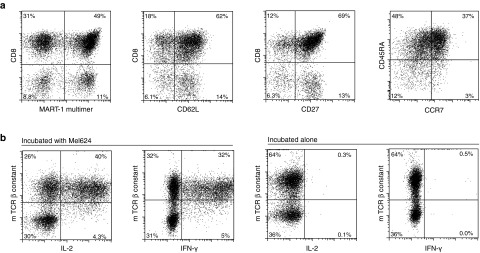
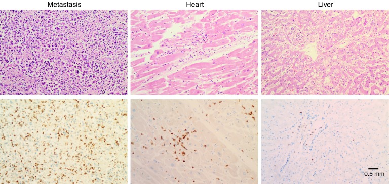
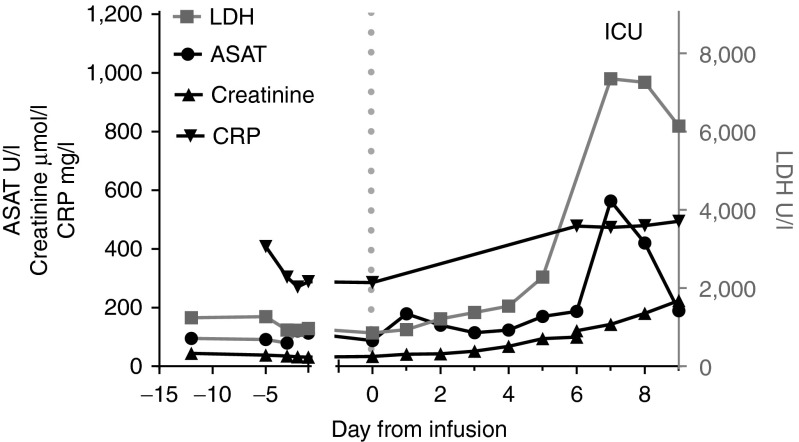
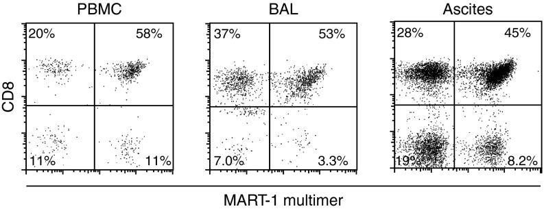
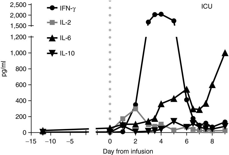
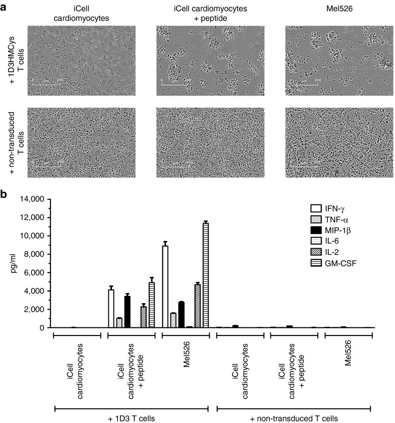
Here, we describe a fatal serious adverse event observed in a patient infused with autologous T-cell receptor (TCR) transduced T cells. This TCR, originally obtained from a melanoma patient, recognizes the well-described HLA-A*0201 restricted 26-35 epitope of MART-1, and was not affinity enhanced. Patient 1 with metastatic melanoma experienced a cerebral hemorrhage, epileptic seizures, and a witnessed cardiac arrest 6 days after cell infusion. Three days later, the patient died from multiple organ failure and irreversible neurologic damage. After T-cell infusion, levels of IL-6, IFN-γ, C-reactive protein (CRP), and procalcitonin increased to extreme levels, indicative of a cytokine release syndrome or T-cell-mediated inflammatory response. Infused T cells could be recovered from blood, broncho-alveolar lavage, ascites, and after autopsy from tumor sites and heart tissue. High levels of NT-proBNP indicate semi-acute heart failure. No cross reactivity of the modified T cells toward a beating cardiomyocyte culture was observed. Together, these observations suggest that high levels of inflammatory cytokines alone or in combination with semi-acute heart failure and epileptic seizure may have contributed substantially to the occurrence of the acute and lethal event. Protocol modifications to limit the risk of T-cell activation-induced toxicity are discussed.


Figures

Figure 1
Figure 1
Characteristics of the final infusion product. (a) Phenotypic analysis of infusion product, gated on propidium iodide negative lymphocytes. (b) Functional analysis of T-cell receptor (TCR)-transduced cells. TCR-transduced cells were cultured alone or incubated with HLA-A2+ MART-1+ Mel624 cells. Intracellular cytokine production was determined after 15 hours of incubation.
Figure 2
Figure 2
Analysis of T-cell infiltration in metastasis, heart, and liver. Upper panel shows H&E staining, lower panel shows CD3 staining.
Figure 3
Figure 3
Blood values during treatment. Dotted line indicates the day of T-cell infusion. Normal reference values are <247 U/l LDH, <31 U/l ASAT, 40–95 µmol/l creatinine, and <8 mg/l for C-reactive protein. ASAH, aspartate aminotransferase; LDH, lactate dehydrogenase.
Figure 4
Figure 4
Flow cytometric analysis of PBMCs, broncho-alveolar lavage (BAL), and ascites samples withdrawn from the patient at day 7 upon T-cell infusion, gated on propidium iodide negative, CD3-positive lymphocytes. PBMC, peripheral blood mononuclear cell.
Figure 5
Figure 5
Cytokine profile in serum samples of patient 1. Only the data point at day -13 was measured in plasma. A previous experiment showed that plasma and serum from the same patient gave the same result. Dotted line indicates time of T-cell infusion.
Figure 6
Figure 6
Lack of reactivity of the T-cell infusion against HLA-A2+ induced-pluripotent stem-cell (iPSC)–derived cardiomyocyte (iPSC-CM) cells, as observed by phase contrast microscopy (a) and cytokine analysis (b).

References

    1. Rosenberg, SA, Yang, JC, Sherry, RM, Kammula, US, Hughes, MS, Phan, GQ et al. (2011). Durable complete responses in heavily pretreated patients with metastatic melanoma using T-cell transfer immunotherapy. Clin Cancer Res 17: 4550–4557. - PMC - PubMed
    1. Morgan, RA, Dudley, ME, Wunderlich, JR, Hughes, MS, Yang, JC, Sherry, RM et al. (2006). Cancer regression in patients after transfer of genetically engineered lymphocytes. Science 314: 126–129. - PMC - PubMed
    1. Johnson, LA, Morgan, RA, Dudley, ME, Cassard, L, Yang, JC, Hughes, MS et al. (2009). Gene therapy with human and mouse T-cell receptors mediates cancer regression and targets normal tissues expressing cognate antigen. Blood 114: 535–546. - PMC - PubMed
    1. Robbins, PF, Morgan, RA, Feldman, SA, Yang, JC, Sherry, RM, Dudley, ME et al. (2011). Tumor regression in patients with metastatic synovial cell sarcoma and melanoma using genetically engineered lymphocytes reactive with NY-ESO-1. J Clin Oncol 29: 917–924. - PMC - PubMed
    1. Chodon, T, Comin-Anduix, B, Chmielowski, B, Koya, RC, Wu, Z, Auerbach, M et al. (2014). Adoptive transfer of MART-1 T-cell receptor transgenic lymphocytes and dendritic cell vaccination in patients with metastatic melanoma. Clin Cancer Res 20: 2457–2465. - PMC - PubMed

Publication types

MeSH terms