Comparative Study
. 2015 Apr 21;20(1):49.
doi: 10.1186/s40001-015-0140-6.

Limitations of mammography in the diagnosis of breast diseases compared with ultrasonography: a single-center retrospective analysis of 274 cases

Affiliations
Comparative Study

Limitations of mammography in the diagnosis of breast diseases compared with ultrasonography: a single-center retrospective analysis of 274 cases

Hong Zhao et al. Eur J Med Res. .

Abstract

Background: The aim of this study is to compare X-ray mammography (MG) and ultrasonography (US) in the diagnosis of breast diseases in Chinese women.

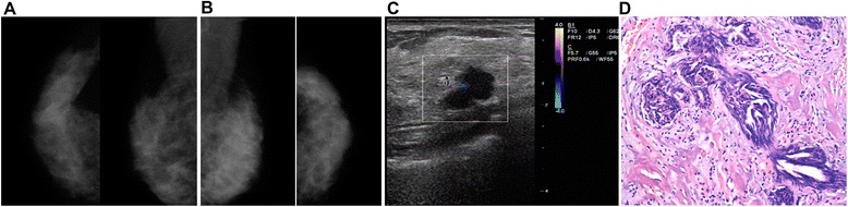
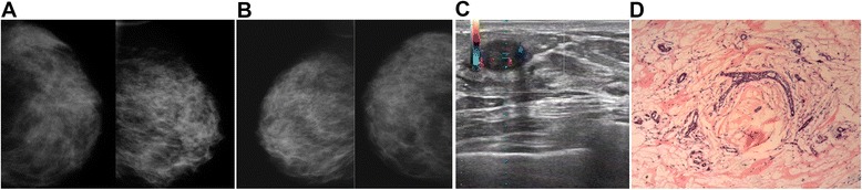
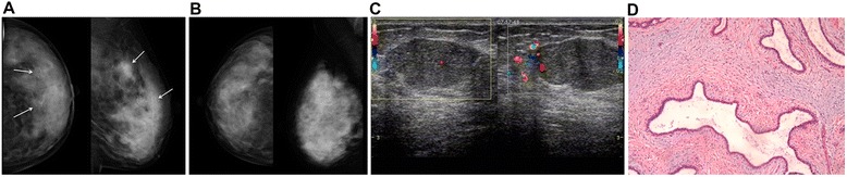
Methods: We retrospectively analyzed X-ray mammograms of 274 patients with US and surgical/pathological results of breast diseases diagnosed at The Second Affiliated Hospital of Anhui Medical University (Hefei, China) between March 2011 and November 2014. The MG and US data were compared to surgical records using the results from post-surgical pathological examinations as the gold standard.

Results: The overall sensitivity, specificity, accuracy, false-positive, false-negative, positive predictive value, and negative predictive value for the detection of breast cancer were 88.5%, 57.9%, 73.7%, 42.1%, 11.5%, 69.2%, and 82.5%, respectively, for MG and 95.9%, 66.7%, 81.8%, 33.3%, 4.1%, 75.5%, and 93.8%, respectively, for US. Of the 274 cases, lesion size by MG agreed with surgery in 133 (48.5%) patients compared with 216 (78.8%) by US (P < 0.01). Lesion location by MG agreed with surgery in 146 (53.3%) patients compared with 257 (93.8%) by US (P < 0.01). These values were then stratified according to age, menstrual status, breast density, and breast volume, and the agreement rates of MG with surgery were lower than that of US (all P < 0.01), except when the lesion size was >5 cm (P > 0.05).

Conclusions: US was better than MG in the preoperative evaluation of breast diseases of Chinese women. These results suggest that US could be more useful for detecting breast lesions in China, especially for younger women with dense breasts.


Figures

Figure 1
Figure 1
MG, US, and post-surgical pathology results from a 43-year-old patient with a lump in her right breast. (A) Craniocaudal and mediolateral oblique views of the molybdenum target MG of the right breast. MG were unsatisfactory (BI-RADS category 0) because of the dense gland structure. (B) MG of the left breast of the same patient. (C) US detected an irregular hypoechoic lesion with clear boundaries. (D) Intra-operative pathology revealed an invasive ductal carcinoma (HE staining, ×100). This is a typical example of a missed diagnosis of breast disease by MG.
Figure 2
Figure 2
MG, US, and post-surgical pathology results from a 46-year-old patient with a lump in her left breast. (A) MG of the left breast could not identify the location of the lesions. (B) MG of the right breast of the same patient. (C) US showed an oval mass in her left breast. (D) Post-surgical pathological examination revealed adenosis of the left breast complicated by fibroadenoma (HE staining, ×100). This represents a typical example of misdiagnosed lesion sites by MG compared with pathological examination.
Figure 3
Figure 3
MG, US, and post-surgical pathology results from a 38-year-old patient with a lump in her left breast. (A) Craniocaudal and mediolateral oblique MG of the left breast displayed lesions in the left outer breast, with multiple clusters of microcalcifications, unclear lesion boundaries (white arrows), and unknown lesion sizes. (B) MG of the right breast of the same patient. (C) The size of lesion was 3 cm by US. (D) Post-surgical pathological examination revealed adenosis of the breast complicated by fibroadenoma, with focal calcifications (HE staining, ×100). This represents a typical example of the inability of MG to correctly determine the lesion boundaries and size compared with pathological examination.

References

    1. Brem RF, Rapelyea JA, Zisman G, Hoffmeister JW, Desimio MP. Evaluation of breast cancer with a computer-aided detection system by mammographic appearance and histopathology. Cancer. 2005;104:931–5. doi: 10.1002/cncr.21255. - DOI - PubMed
    1. Boyd NF, Guo H, Martin LJ, Sun L, Stone J, Fishell E, et al. Mammographic density and the risk and detection of breast cancer. N Engl J Med. 2007;356:227–36. doi: 10.1056/NEJMoa062790. - DOI - PubMed
    1. Maskarinec G, Meng L, Ursin G. Ethnic differences in mammographic densities. Int J Epidemiol. 2001;30:959–65. doi: 10.1093/ije/30.5.959. - DOI - PubMed
    1. Pisano ED, Gatsonis C, Hendrick E, Yaffe M, Baum JK, Acharyya S, et al. Diagnostic performance of digital versus film mammography for breast-cancer screening. N Engl J Med. 2005;353:1773–83. doi: 10.1056/NEJMoa052911. - DOI - PubMed
    1. Shao H, Li B, Zhang X, Xiong Z, Liu Y, Tang G. Comparison of the diagnostic efficiency for breast cancer in Chinese women using mammography, ultrasound, MRI, and different combinations of these imaging modalities. J Xray Sci Technol. 2013;21:283–92. - PubMed

Publication types