

Initial Evaluation of [(18)F]DCFPyL for Prostate-Specific Membrane Antigen (PSMA)-Targeted PET Imaging of Prostate Cancer
- PMID: 25896814
- PMCID: PMC4531836
- DOI: 10.1007/s11307-015-0850-8
Initial Evaluation of [(18)F]DCFPyL for Prostate-Specific Membrane Antigen (PSMA)-Targeted PET Imaging of Prostate Cancer
Abstract
Purpose: Prostate-specific membrane antigen (PSMA) is a recognized target for imaging prostate cancer. Here we present initial safety, biodistribution, and radiation dosimetry results with [(18)F]DCFPyL, a second-generation fluorine-18-labeled small-molecule PSMA inhibitor, in patients with prostate cancer.
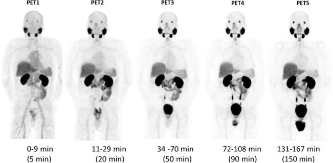
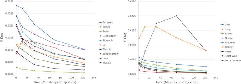
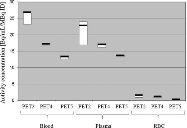
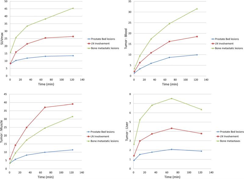
Procedures: Biodistribution was evaluated using sequential positron-emission tomography (PET) scans in nine patients with prostate cancer. Time-activity curves from the most avid tumor foci were determined. The radiation dose to selected organs was estimated using OLINDA/EXM.
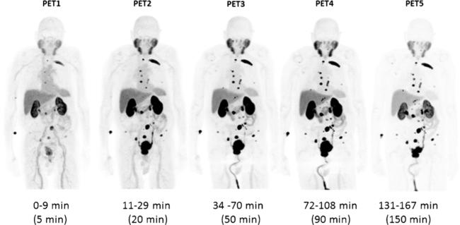
Results: No major radiotracer-specific adverse events were observed. Physiologic accumulation was observed in known sites of PSMA expression. Accumulation in putative sites of prostate cancer was observed (SUVmax up to >100, and tumor-to-blood ratios up to >50). The effective radiation dose from [(18)F]DCFPyL was 0.0139 mGy/MBq or 5 mGy (0.5 rem) from an injected dose of 370 MBq (10 mCi).
Conclusions: [(18)F]DCFPyL is safe with biodistribution as expected, and its accumulation is high in presumed primary and metastatic foci. The radiation dose from [(18)F]DCFPyL is similar to that from other PET radiotracers.
Figures
References
-
- Siegel R, Ma J, Zou Z, Jemal A. Cancer statistics. CA Cancer J Clin. 2014;64:9–29. - PubMed
-
- Shinohara K, Wheeler TM, Scardino PT. The appearance of prostate cancer on transrectal ultrasonography: correlation of imaging and pathological examinations. J Urol. 1989;142:76–82. - PubMed
-
- Hricak H, Dooms GC, Jeffrey RB, et al. Prostatic carcinoma: staging by clinical assessment, CT, and MR imaging. Radiology. 1987;162:331–336. - PubMed
-
- Scheidler J, Hricak H, Vigneron DB, et al. Prostate cancer: localization with three-dimensional proton MR spectroscopic imaging—clinicopathologic study. Radiology. 1999;213:473–480. - PubMed
-
- Blomqvist L, Carlsson S, Gjertsson P, et al. Limited evidence for the use of imaging to detect prostate cancer: a systematic review. Eur J Radiol. 2014;83:1601–1606. - PubMed
Publication types
MeSH terms
Substances
Grants and funding
- R01 CA184228/CA/NCI NIH HHS/United States
- R01 CA116477/CA/NCI NIH HHS/United States
- R01 CA134675/CA/NCI NIH HHS/United States
- UL1 TR001079/TR/NCATS NIH HHS/United States
- P30 CA006973/CA/NCI NIH HHS/United States
- CA134675/CA/NCI NIH HHS/United States
- CA183031/CA/NCI NIH HHS/United States
- CA103175/CA/NCI NIH HHS/United States
- P50 CA103175/CA/NCI NIH HHS/United States
- EB006351/EB/NIBIB NIH HHS/United States
- U01 CA183031/CA/NCI NIH HHS/United States
- T32 EB006351/EB/NIBIB NIH HHS/United States
- CA184228/CA/NCI NIH HHS/United States
LinkOut - more resources
Full Text Sources
Other Literature Sources
Medical
Miscellaneous
