

Case Reports
doi: 10.1186/s12957-015-0581-y.
Posterior mediastinal ectopic meningioma: a case report
Affiliations
- PMID: 25896915
- PMCID: PMC4417514
- DOI: 10.1186/s12957-015-0581-y
Item in Clipboard
Case Reports
Posterior mediastinal ectopic meningioma: a case report
World J Surg Oncol.
.
Abstract
Primary ectopic meningiomas occurring in the mediastinal region are extremely rare. So far, only five cases of primary mediastinal meningioma have been reported in the literatures. The imaging characteristics and the clinicopathological significance of mediastinal psammomatous meningioma have not been detailed. Here, we report the case of a 42-year-old male with primary posterior mediastinal psammomatous meningioma. The clinical features, imaging, and pathological findings are carefully analyzed, and the relevant literatures were reviewed.
Figures

A chest lateral radiograph shows a large mass with lots of calcification in the mediastinum; esophagus barium opacification shows that the middle and inferior segment of the esophagus was compressed by the mass.
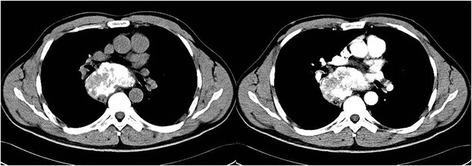
Chest unenhanced CT scan shows a heterogeneous mass with calcification located in the posterior mediastinum. Its border was clearly demarcated, and enhanced CT scan shows the mass with little enhancement.

Axial T1-weighted MRI shows a well-circumscribed isointense-hypointense mass located in the posterior mediastinum, and coronal T2-weighted MRI shows a hypointense tumor located under the tracheal carina.

Hematoxylin and eosin (H-E) staining shows that the tumor is composed prominently of elongated spindle cells and collagen fibers; numerous typical whorl formations and multiple psammoma bodies are observed in the tumor. Immunohistochemistry shows positive staining for epithelial membrane antigen (EMA). (Magnification shown at × 100).
References
Publication types
MeSH terms
LinkOut - more resources
Full Text Sources
Other Literature Sources
