. 2015 Aug 1;21(15):3501-11.
doi: 10.1158/1078-0432.CCR-14-3141. Epub 2015 Apr 20.

Clinically Relevant Molecular Subtypes in Leiomyosarcoma

Affiliations

Clinically Relevant Molecular Subtypes in Leiomyosarcoma

Xiangqian Guo et al. Clin Cancer Res. .

Abstract

Purpose: Leiomyosarcoma is a malignant neoplasm with smooth muscle differentiation. Little is known about its molecular heterogeneity and no targeted therapy currently exists for leiomyosarcoma. Recognition of different molecular subtypes is necessary to evaluate novel therapeutic options. In a previous study on 51 leiomyosarcomas, we identified three molecular subtypes in leiomyosarcoma. The current study was performed to determine whether the existence of these subtypes could be confirmed in independent cohorts.

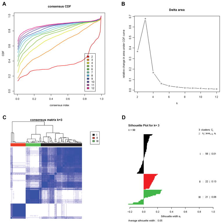
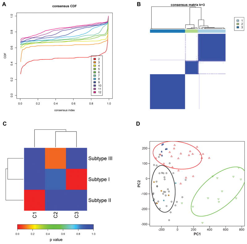
Experimental design: Ninety-nine cases of leiomyosarcoma were expression profiled with 3'end RNA-Sequencing (3SEQ). Consensus clustering was conducted to determine the optimal number of subtypes.

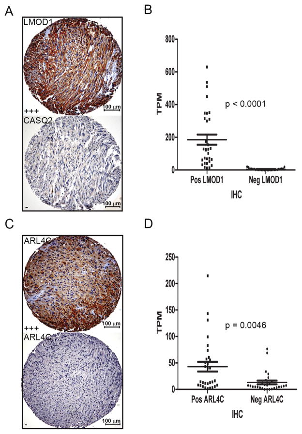
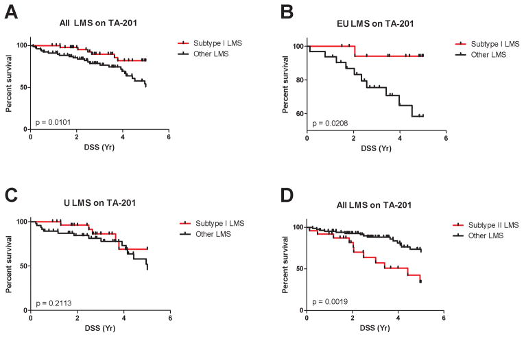
Results: We identified 3 leiomyosarcoma molecular subtypes and confirmed this finding by analyzing publically available data on 82 leiomyosarcoma from The Cancer Genome Atlas (TCGA). We identified two new formalin-fixed, paraffin-embedded tissue-compatible diagnostic immunohistochemical markers; LMOD1 for subtype I leiomyosarcoma and ARL4C for subtype II leiomyosarcoma. A leiomyosarcoma tissue microarray with known clinical outcome was used to show that subtype I leiomyosarcoma is associated with good outcome in extrauterine leiomyosarcoma while subtype II leiomyosarcoma is associated with poor prognosis in both uterine and extrauterine leiomyosarcoma. The leiomyosarcoma subtypes showed significant differences in expression levels for genes for which novel targeted therapies are being developed, suggesting that leiomyosarcoma subtypes may respond differentially to these targeted therapies.

Conclusions: We confirm the existence of 3 molecular subtypes in leiomyosarcoma using two independent datasets and show that the different molecular subtypes are associated with distinct clinical outcomes. The findings offer an opportunity for treating leiomyosarcoma in a subtype-specific targeted approach.


Conflict of interest statement

Disclosure of Potential Conflicts of Interest

No potential conflicts of interest were disclosed.

Figures

Figure 1
Figure 1. Identification of three molecular subtypes in LMS
A). Empirical cumulative distribution plots as a function of the number of molecular subtypes in 3SEQ cohort. B). Relative increase in the area under the CDF curve with increasing number of molecular subtypes. C). Consensus clustering matrix of LMS samples using 3 molecular subtypes. D). Silhouette analysis of LMS samples based on the assignments from Consensus Clustering.
Figure 2
Figure 2. Identification of molecular subtypes of LMS in TCGA data
A). Empirical cumulative distribution plots as a function of the number of molecular subtypes in TCGA cohort. B). Consensus clustering matrix of LMS samples using 3 molecular subtypes. C). Subclass association (SA) matrix between 3SEQ data (subtype I, subtype II, subtype III) and TCGA data (subtype C1, subtype C2, subtype C3). p value is corrected with FDR. D). Principal component analysis (PCA) of 70 core 3SEQ LMS cases with other diagnoses. Subtype I LMS-black circle; subtype II LMS-red triangle: subtype III LMS-green triangle; leiomyoma-cyan solid circle; myometrium-orange solid circle; UPS-blue solid pyramid. The elliptical contours represent the normal probability of 0.75.
Figure 3
Figure 3. Immunohistochemical markers for subtype I and subtype II LMS
A). Representative staining for LMOD1 (Score +++) and CASQ2 (Negative) for a subtype I LMS case (case # 10129, scale bar = 100μm, 10X). B). Comparison of 3SEQ mRNA quantification and immunostaining results on TA-381 that contains 58 cases of LMS analyzed by 3SEQ; LMS cases positive for anti-LMOD1 staining have significantly higher mRNA expression levels (TPM) than LMS cases negative for anti-LMOD1 (p < 0.0001). The p value was derived from unpaired t test with Welch’s correction. C). Positive and negative staining by ARL4C antibody for representative subtype II LMS (top, case # 10019) and subtype I LMS (bottom case # 9994, scale bar = 100μm, 10X). D). LMS cases positive for anti-ARL4C staining have significantly higher mRNA expression levels (TPM) than LMS cases negative for anti-ARL4C (p = 0.0046).
Figure 4
Figure 4. Immunohistochemical markers for subtype I and subtype II LMS predict different clinical outcomes
TA-201 with 127 cases of LMS with known clinical outcome was stained for the panel of 5 subtype I LMS markers and the ARL4C marker for subtype II LMS. The p value was from Log-rank (Mantel-Cox) test. Coordinate expression of subtype I biomarkers was associated with good outcome when analysed on all cases (panel A) but this was mainly due to an effect on extra-uterine (EU) LMS (panel B) but not uterine (U) LMS (panel C). Subtype II biomarker ARL4C predicted worse outcome in LMS (panel D), with equal effect on extra-uterine and uterine LMS (not shown).

References

    1. Gustafson P, Willen H, Baldetorp B, Ferno M, Akerman M, Rydholm A. Soft tissue leiomyosarcoma. A population-based epidemiologic and prognostic study of 48 patients, including cellular DNA content. Cancer. 1992;70:114–9. - PubMed
    1. Toro JR, Travis LB, Wu HJ, Zhu K, Fletcher CD, Devesa SS. Incidence patterns of soft tissue sarcomas, regardless of primary site, in the surveillance, epidemiology and end results program, 1978–2001: An analysis of 26,758 cases. Int J Cancer. 2006;119:2922–30. - PubMed
    1. Witt H, Mack SC, Ryzhova M, Bender S, Sill M, Isserlin R, et al. Delineation of two clinically and molecularly distinct subgroups of posterior fossa ependymoma. Cancer Cell. 2011;20:143–57. - PMC - PubMed
    1. Marisa L, de Reynies A, Duval A, Selves J, Gaub MP, Vescovo L, et al. Gene expression classification of colon cancer into molecular subtypes: characterization, validation, and prognostic value. PLoS Med. 2013;10:e1001453. - PMC - PubMed
    1. Lapointe J, Li C, Higgins JP, van de Rijn M, Bair E, Montgomery K, et al. Gene expression profiling identifies clinically relevant subtypes of prostate cancer. Proc Natl Acad Sci U S A. 2004;101:811–6. - PMC - PubMed

Publication types

MeSH terms