

Quantitative optical coherence tomography angiography of vascular abnormalities in the living human eye
- PMID: 25897021
- PMCID: PMC4426471
- DOI: 10.1073/pnas.1500185112
Quantitative optical coherence tomography angiography of vascular abnormalities in the living human eye
Abstract
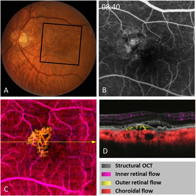
Retinal vascular diseases are important causes of vision loss. A detailed evaluation of the vascular abnormalities facilitates diagnosis and treatment in these diseases. Optical coherence tomography (OCT) angiography using the highly efficient split-spectrum amplitude decorrelation angiography algorithm offers an alternative to conventional dye-based retinal angiography. OCT angiography has several advantages, including 3D visualization of retinal and choroidal circulations (including the choriocapillaris) and avoidance of dye injection-related complications. Results from six illustrative cases are reported. In diabetic retinopathy, OCT angiography can detect neovascularization and quantify ischemia. In age-related macular degeneration, choroidal neovascularization can be observed without the obscuration of details caused by dye leakage in conventional angiography. Choriocapillaris dysfunction can be detected in the nonneovascular form of the disease, furthering our understanding of pathogenesis. In choroideremia, OCT's ability to show choroidal and retinal vascular dysfunction separately may be valuable in predicting progression and assessing treatment response. OCT angiography shows promise as a noninvasive alternative to dye-based angiography for highly detailed, in vivo, 3D, quantitative evaluation of retinal vascular abnormalities.
Keywords: ocular circulation; ophthalmic imaging; optical coherence tomography angiography.
Conflict of interest statement
Conflict of interest statement: Oregon Health & Science University (OHSU), Y.J., J.G.F., and D.H. have a significant financial interest in Optovue, Inc., a company that may have a commercial interest in the results of this research and technology. These potential conflicts of interest have been reviewed and managed by OHSU. J.G.F. and D.H. receive royalties on an optical coherence tomography patent licensed by the Massachusetts Institute of Technology (MIT) to Carl Zeiss Meditec. J.G.F. and J.H. receive royalties from intellectual property owned by MIT and licensed to Optovue, Inc. Other authors do not have financial interest in the subject of this article.
Figures


References
-
- Congdon N, et al. Eye Diseases Prevalence Research Group Causes and prevalence of visual impairment among adults in the United States. Arch Ophthalmol. 2004;122(4):477–485. - PubMed
-
- López-Sáez MP, et al. Fluorescein-induced allergic reaction. Ann Allergy Asthma Immunol. 1998;81(5 Pt 1):428–430. - PubMed
-
- Wang RK, et al. Three dimensional optical angiography. Opt Express. 2007;15(7):4083–4097. - PubMed
-
- Grulkowski I, et al. Scanning protocols dedicated to smart velocity ranging in spectral OCT. Opt Express. 2009;17(26):23736–23754. - PubMed
Publication types
MeSH terms
Substances
Grants and funding
- R01 EY023285/EY/NEI NIH HHS/United States
- P30 EY010572/EY/NEI NIH HHS/United States
- UL1 TR000128/TR/NCATS NIH HHS/United States
- UL1TR000128/TR/NCATS NIH HHS/United States
- P30-EY010572/EY/NEI NIH HHS/United States
- K08 EY021186/EY/NEI NIH HHS/United States
- R01-EY024544/EY/NEI NIH HHS/United States
- DP3 DK104397/DK/NIDDK NIH HHS/United States
- R01-EY11289/EY/NEI NIH HHS/United States
- R01 EY024544/EY/NEI NIH HHS/United States
- T32-EY23211/EY/NEI NIH HHS/United States
- K08-EY021186/EY/NEI NIH HHS/United States
- R01 EY011289/EY/NEI NIH HHS/United States
- R01-EY023285/EY/NEI NIH HHS/United States
- T32 EY023211/EY/NEI NIH HHS/United States
LinkOut - more resources
Full Text Sources
Other Literature Sources
Medical
