. 2015 May 29;290(22):14218-25.
doi: 10.1074/jbc.M115.650762. Epub 2015 Apr 20.

The Association of the Xeroderma Pigmentosum Group D DNA Helicase (XPD) with Transcription Factor IIH Is Regulated by the Cytosolic Iron-Sulfur Cluster Assembly Pathway

Affiliations

The Association of the Xeroderma Pigmentosum Group D DNA Helicase (XPD) with Transcription Factor IIH Is Regulated by the Cytosolic Iron-Sulfur Cluster Assembly Pathway

Ajay A Vashisht et al. J Biol Chem. .

Abstract

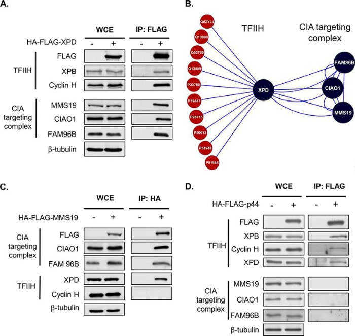
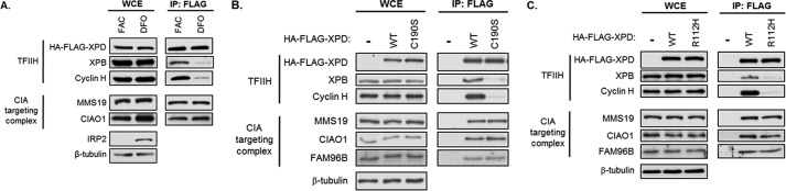
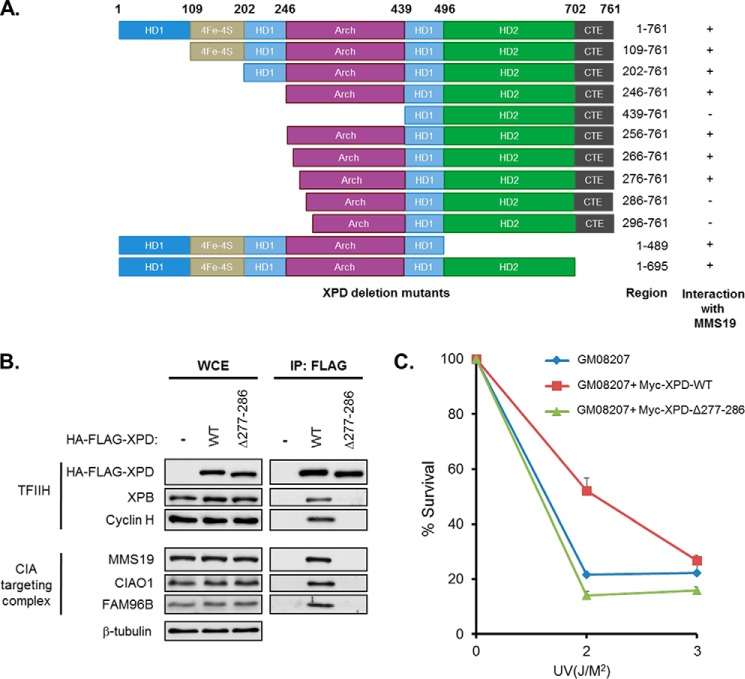
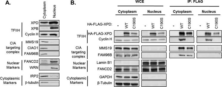
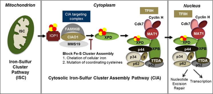
Xeroderma pigmentosum group D (XPD) helicase is a component of the transcription factor IIH (TFIIH) transcription complex and plays essential roles in transcription and nucleotide excision repair. Although iron-sulfur (Fe-S) cluster binding by XPD is required for activity, the process mediating Fe-S cluster assembly remains poorly understood. We recently identified a cytoplasmic Fe-S cluster assembly (CIA) targeting complex composed of MMS19, CIAO1, and FAM96B that is required for the biogenesis of extramitochondrial Fe-S proteins including XPD. Here, we use XPD as a prototypical Fe-S protein to further characterize how Fe-S assembly is facilitated by the CIA targeting complex. Multiple lines of evidence indicate that this process occurs in a stepwise fashion in which XPD acquires a Fe-S cluster from the CIA targeting complex before assembling into TFIIH. First, XPD was found to associate in a mutually exclusive fashion with either TFIIH or the CIA targeting complex. Second, disrupting Fe-S cluster assembly on XPD by either 1) depleting cellular iron levels or 2) utilizing XPD mutants defective in either Fe-S cluster or CIA targeting complex binding blocks Fe-S cluster assembly and prevents XPD incorporation into TFIIH. Finally, subcellular fractionation studies indicate that the association of XPD with the CIA targeting complex occurs in the cytoplasm, whereas its association with TFIIH occurs largely in the nucleus where TFIIH functions. Together, these data establish a sequential assembly process for Fe-S assembly on XPD and highlight the existence of quality control mechanisms that prevent the incorporation of immature apoproteins into their cellular complexes.

Keywords: DNA helicase; DNA repair; iron metabolism; iron-sulfur protein; proteomics.


Figures

FIGURE 1.
FIGURE 1.
Human XPD associates with TFIIH and the CIA targeting complex in a mutually exclusive manner. A, Flp-InTM TRExTM-293-derived stable cells lines expressing HA-FLAG-XPD were induced with doxycycline (500 ng/ml). XPD was immunopurified using FLAG antibodies, and samples were analyzed by SDS-PAGE and blotted with indicated antibodies. WCE, whole cell extracts. B, schematic summarizing protein interactions identified by proteomic mass spectrometry between XPD, TFIIH components, and the CIA targeting complex. A complete list of the TFIIH and CIA targeting complex components identified in XPD proteomic analysis as well as all identified XPD-interacting proteins can be found in Table 1 and supplemental Table 1, respectively. C and D, Flp-InTM TRExTM-293-derived stable cell lines expressing HA-FLAG-MMS19 (C) or HA-FLAG-p44 (D) were induced overnight with doxycycline (500 ng/ml), and HA-FLAG-tagged proteins were immunoprecipitated with FLAG antibodies (HA-FLAG-MMS19 and HA-FLAG-p44). Whole cell extracts and immunoprecipitates (IP) were immunoblotted with the indicated antibodies. The parental Flp-InTM TRExTM-293 cell line was used as a control in all cases.
FIGURE 2.
FIGURE 2.
Incorporation of XPD into TFIIH requires both adequate cellular iron levels and the ability of XPD to coordinate an Fe-S cluster. A, Flp-InTM TRExTM-293 cells stably expressing 3HA-3FLAG-XPD or control cells were induced overnight with doxycycline (500 ng/ml) and treated for 8 h with 100 μg/ml ferric ammonium citrate (FAC; Thermo Fisher Scientific) or 100 μm desferrioxamine mesylate (DFO; Sigma). HA-FLAG-XPD was immunoprecipitated with FLAG antibody. Whole cell extract (WCE) and immunoprecipitates (IP) were probed with the indicated antibodies. B and C, Flp-InTM TRExTM-293 derived cell lines stably expressing HA-FLAG-XPD-WT, HA-FLAG-XPD-C190S, or HA-FLAG-XPD-R112H were induced overnight with doxycycline (500 ng/ml). Cell lysates were prepared, and HA-FLAG-XPD wild type and mutant proteins were immunoprecipitated using FLAG antibody. WCE and immunoprecipitates were blotted with indicated antibodies.
FIGURE 3.
FIGURE 3.
XPD associates with MMS19 via a peptide docking site that is indispensable for its assembly into TFIIH and its DNA repair functions. A, summary of protein-protein interaction studies performed using co-immunoprecipitation assays between a series of XPD deletion mutants and MMS19. B, Flp-InTM TRExTM-293 cells stably expressing HA-FLAG-XPD-WT, HA-FLAG-XPD-Δ277–286, or control cells were induced overnight with doxycycline (500 ng/ml). Whole cell extracts (WCE) and immunoprecipitates (IP) using FLAG antibody were probed with the indicated antibodies. C, XPD patient fibroblast cells (GM08207) were obtained from Coriell Institute for Medical Research and complemented with XPD-WT and XPD-Δ277–286 (defective in MMS19 binding) and were tested for their sensitivity to UV irradiation. The number of viable cells was counted for each set after 7 days of UV treatment, and % survival rate was plotted as mean ± S.E. (n = 3).
FIGURE 4.
FIGURE 4.
Subcellular distribution of XPD, TFIIH, and the CIA targeting complex. A, nuclear and cytoplasmic fractions were prepared from HeLa cells and immunoblotted with antibodies against TFIIH components (XPD, XPB, and cyclin H), CIA targeting complex proteins (MMS19, CIAO1, and FAM96B), and nuclear (WRN) and cytosolic (iron regulatory protein 2 (IRP2) markers. B, HA-FLAG-XPD-WT was immunoprecipitated using FLAG beads from nuclear and cytoplasmic fractions prepared from Flp-InTM TRExTM-HeLa cells stably expressing HA-FLAG-XPD-WT after overnight induction with doxycycline (500 ng/ml). WCE and immunoprecipitates (IP) were blotted with the indicated antibodies.
FIGURE 5.
FIGURE 5.
Fe-S cluster assembly of XPD is required for its integration into TFIIH. Extramitochondrial Fe-S cluster biogenesis depends on the activity of the mitochondrially localized iron-sulfur cluster (ISC) biogenesis machinery, which generates an as of yet unidentified sulfur-based compound that is exported to the cytosol (5). The cytoplasmic iron-sulfur cluster assembly (CIA pathway) then catalyzes the assembly of Fe-S clusters on cytoplasmic protein scaffolds, which are subsequently transferred to apoproteins in a manner dependent on the iron-only hydrogenase IOP1 and the CIA targeting complex (MMS19, CIAO1, and FAM96B) (12). Our data suggest that Fe-S cluster delivery to apoproteins such as XPD occurs in the cytoplasm via a stepwise pathway in which XPD is first recruited to the CIA machinery via direct protein-protein interactions. XPD then remains stably associated with the CIA targeting complex until cluster transfer is completed, which then triggers its release and subsequent assembly into TFIIH. Once TFIIH is properly assembled, it translocates into the nucleus where it performs its essential functions in transcription and DNA repair. Inhibiting Fe-S cluster assembly during this process results in the sequestration of XPD by the CIA targeting complex preventing the incorporation of apoXPD into TFIIH.

Similar articles

Cited by

References

    1. Friedberg E. C. (2001) How nucleotide excision repair protects against cancer. Nat. Rev. Cancer 1, 22–33 - PubMed
    1. Lehmann A. R. (2001) The xeroderma pigmentosum group D (XPD) gene: one gene, two functions, three diseases. Genes Dev. 15, 15–23 - PubMed
    1. Netz D. J., Mascarenhas J., Stehling O., Pierik A. J., Lill R. (2014) Maturation of cytosolic and nuclear iron-sulfur proteins. Trends Cell Biol. 24, 303–312 - PubMed
    1. Ye H., Rouault T. A. (2010) Human iron-sulfur cluster assembly, cellular iron homeostasis, and disease. Biochemistry 49, 4945–4956 - PMC - PubMed
    1. Lill R. (2009) Function and biogenesis of iron-sulphur proteins. Nature 460, 831–838 - PubMed

Publication types