

doi: 10.1093/nar/gkv366.
Epub 2015 Apr 19.
Single-cell, locus-specific bisulfite sequencing (SLBS) for direct detection of epimutations in DNA methylation patterns
Affiliations
- PMID: 25897117
- PMCID: PMC4538804
- DOI: 10.1093/nar/gkv366
Item in Clipboard
Single-cell, locus-specific bisulfite sequencing (SLBS) for direct detection of epimutations in DNA methylation patterns
Nucleic Acids Res.
.
Abstract
Stochastic epigenetic changes drive biological processes, such as development, aging and disease. Yet, epigenetic information is typically collected from millions of cells, thereby precluding a more precise understanding of cell-to-cell variability and the pathogenic history of epimutations. Here we present a novel procedure for directly detecting epimutations in DNA methylation patterns using single-cell, locus-specific bisulfite sequencing (SLBS). We show that within gene promoter regions of mouse hepatocytes the epimutation rate is two orders of magnitude higher than the mutation rate.
© The Author(s) 2015. Published by Oxford University Press on behalf of Nucleic Acids Research.
Figures
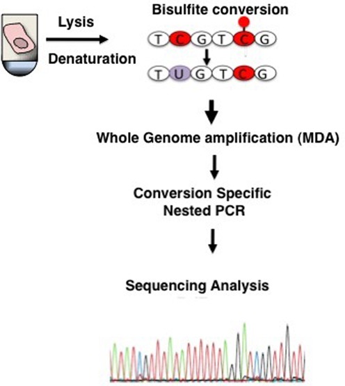
Schematic depiction of single-cell DNA methylation analysis. Single cells are lysed and treated with bisulfite to convert unmethylated cytosines into uracil. After bisulfite treatment, DNA is immediately whole-genome amplified by MDA. DNA methylation patterns are analyzed in a locus-specific way using PCR amplification with primers specific for cytosine-converted DNA.
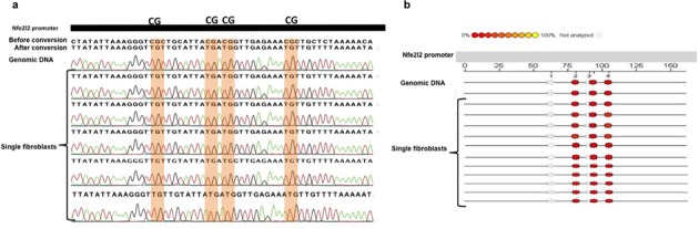
(a) DNA methylation profile of a 180 bp fragment of the Nfe2l2 promoter in single fibroblasts analyzed using Sanger Sequencing. (b) DNA methylation profile of a 180 bp fragment of the Nfe2l2 promoter in single fibroblasts analyzed using Sequenom's EpiTYPER System. The EpiGram is a graphical representation of methylation ratios found in each sample for the amplicon studied. Each sample's nucleotide sequence is displayed as a series of individual CpGs, which are color-coded columns on the same line. The color within the column denotes the level of methylation found at this particular site in the selected sample. The color spectrum ranges from red (0% methylated) CpG units to yellow (100% methylated) CpG units. Gray dots denote not analyzable CpG sites.
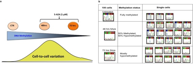
(a) A schematic depiction of the 5-Aza-time course experiment in single fibroblasts. (b) Representative example of DNA methylation profiles of Oct4-specific CpG sites in MEFs treated with 1 μM 5-Aza for 48 and 72 h. As expected, Oct4 CpG sites in the untreated single fibroblasts as well as in the 100-cell control were mostly methylated (top panel), while after 72 h of treatment the Oct4 promoter was mostly demethylated in the 100-cell control as well in the single fibroblasts (lower panel). The recovery of both C and T alleles, particularly evident after 48 h of treatment (middle panel), could be interpreted as a 5-Aza-induced hemi-demethylation, caused by incomplete demethylation possibly due to failure of the covalent 5-Aza/Dnmt1 binding on one allele.
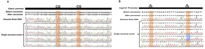
(a) DNA methylation profiling of Gabra1 in single neuronal nuclei. Cytosines of CpG dinucleotides are highlighted in orange. (b) Cyp71a promoter in single neuronal nuclei. Cytosines of CpG dinucleotides are highlighted in orange. Epimutations are highlighted in blue.
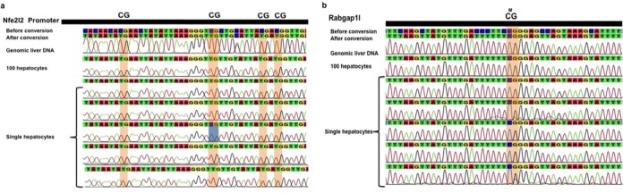
(a) DNA methylation profile of the Nfe2l2 promoter in single hepatocytes. Cytosines of CpG dinucleotides are highlighted in orange. (b) DNA methylation profile of Rabgap1l (intragenic region) in single hepatocytes. Cytosines of CpG dinucleotides are highlighted in orange. Epimutations are highlighted in blue.
References
Publication types
MeSH terms
Substances
Grants and funding
LinkOut - more resources
Full Text Sources
Other Literature Sources
Molecular Biology Databases
