. 2015 May 19;43(9):4758-73.
doi: 10.1093/nar/gkv321. Epub 2015 Apr 19.

A variable DNA recognition site organization establishes the LiaR-mediated cell envelope stress response of enterococci to daptomycin

Affiliations

A variable DNA recognition site organization establishes the LiaR-mediated cell envelope stress response of enterococci to daptomycin

Milya Davlieva et al. Nucleic Acids Res. .

Abstract

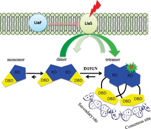
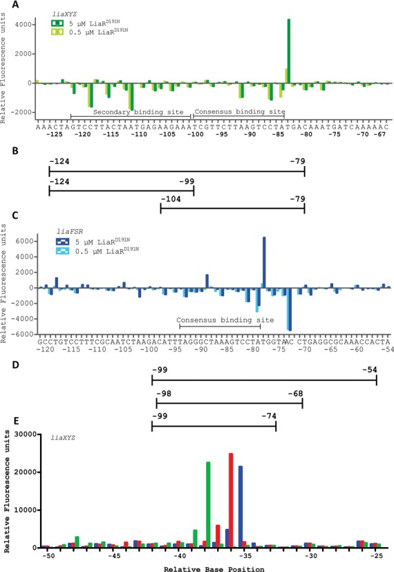
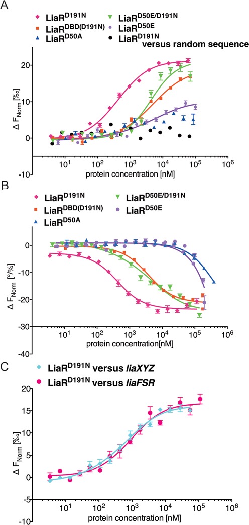
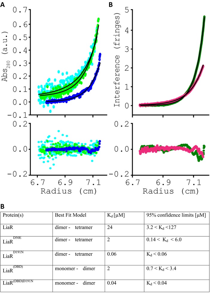
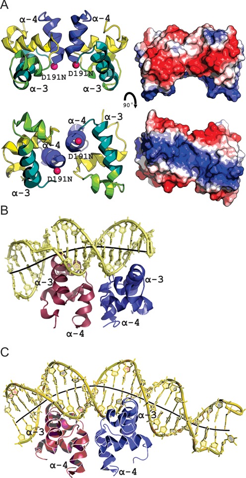
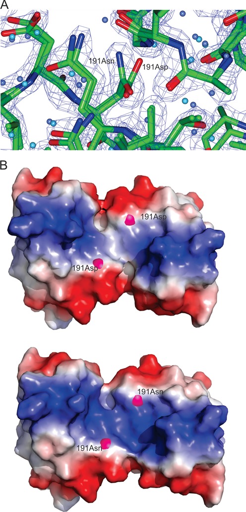
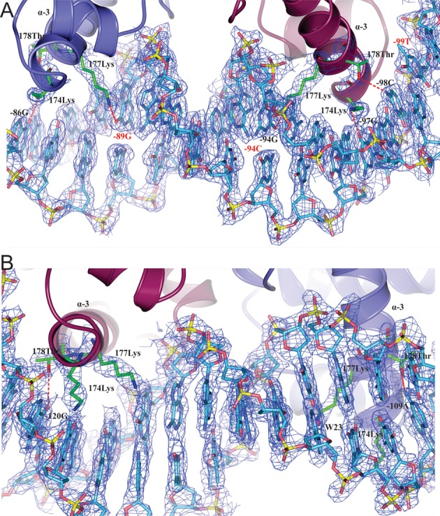
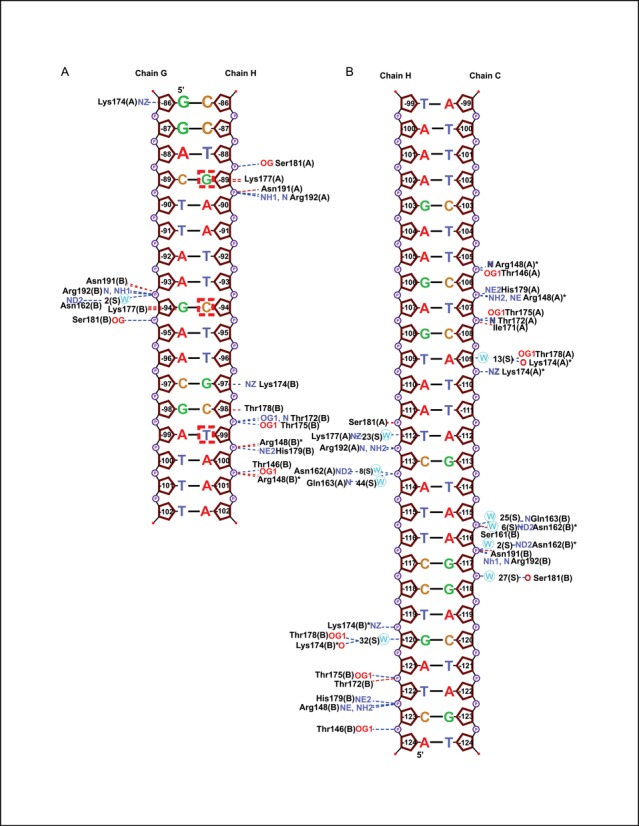
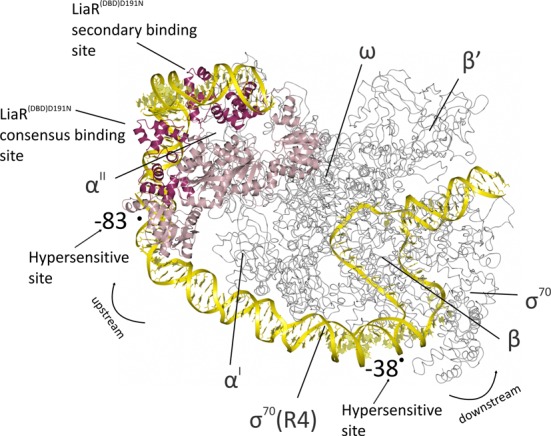
LiaR is a 'master regulator' of the cell envelope stress response in enterococci and many other Gram-positive organisms. Mutations to liaR can lead to antibiotic resistance to a variety of antibiotics including the cyclic lipopeptide daptomycin. LiaR is phosphorylated in response to membrane stress to regulate downstream target operons. Using DNA footprinting of the regions upstream of the liaXYZ and liaFSR operons we show that LiaR binds an extended stretch of DNA that extends beyond the proposed canonical consensus sequence suggesting a more complex level of regulatory control of target operons. We go on to determine the biochemical and structural basis for increased resistance to daptomycin by the adaptive mutation to LiaR (D191N) first identified from the pathogen Enterococcus faecalis S613. LiaR(D191N) increases oligomerization of LiaR to form a constitutively activated tetramer that has high affinity for DNA even in the absence of phosphorylation leading to increased resistance. Crystal structures of the LiaR DNA binding domain complexed to the putative consensus sequence as well as an adjoining secondary sequence show that upon binding, LiaR induces DNA bending that is consistent with increased recruitment of RNA polymerase to the transcription start site and upregulation of target operons.


Figures

Figure 1.
Figure 1.
LiaFSR signaling pathway. In the absence of cell membrane-acting antibiotics, the three-component regulatory system, LiaFSR, is turned ‘OFF’ by the negative interaction of LiaF with LiaS. LiaS responds to membrane stress by phosphorylation of LiaR leading to downstream changes in the transcription of several operons to affect membrane homeostasis. E. faecalis LiaR is present largely as a dimer at physiologically relevant concentrations. Phosphorylation, or mutation to a constitutively active analog like LiaRD191N, induces changes in the conformation of the receiver domain leading to release of DNA binding domain and promoting a self-dimerization event to form an active tetramer able to bind extended DNA sequences. Abbreviations: RD, receiver domain; DBD, DNA binding domain.
Figure 2.
Figure 2.
LiaR binds to extended region of DNA that includes sequences outside the proposed canonical consensus sequence. DNase I Footprinting followed by DFACE was used to identify the DNA sequences protected by LiaRD191N within the upstream regions of the liaXYZ (−320 to +30) and liaFSR (−367 to +30) operons of E. faecalis S613. Protection studies were performed at 0.5 and 5 μM LiaRD191N. DNaseI digestion patterns are shown as histograms (A, C, E) where negative changes in relative fluorescence units indicate regions of protection and positives changes indicate hypersensitivity. All DNaseI sensitivity data are relative to a no protein negative control. (A) LiaRD191N binding to the promoter region of the liaXYZ operon. Nucleotide positions refer to the region of LiaR binding (-120 to -77) on the DNA relative to the translation start site of LiaX. (B/D) Oligonucleotides used for DNA binding studies. (C) LiaRD191N binding to the promoter region of the liaFRS operon. Nucleotide positions refer to the region of LiaR binding (−97 to -68) on the DNA relative to the translation start site of LiaF. The AC bar indicates a compression artifact of two bases during electrophoresis seen in both the fragment pattern and the sequencing reaction results. (E) Superimposed electropherograms showing the shift of a hypersensitive location in the upstream region of the liaXYZ operon in response to increasing concentrations of LiaR (blue, 0 μM; red, 0.5 μM; green 5.0 μM). The hypersensitive position shifts from −35(T) to −36(C) to −38(C) in response to increasing protein concentration.
Figure 3.
Figure 3.
Adaptive mutant LiaRD191N that confers increased daptomycin resistance in E. faecalis S613 dramatically increases LiaR affinity for target DNA sequences. E. faecalis response regulator LiaR–DNA interactions were measured with MST. To determine the Kd, increasing concentrations of LiaRD191N was added to 40 nM of fluorescently labeled DNAs (Supplementary Information). Fnorm (normalized fluorescence) was plotted on the y-axis in per mil [‰] unit (meaning every 1000) against the total concentration of the titrated partner on a log10 scale on the x-axis (21). The resulting Kd values based on average from six independent MST measurements. Note, when the markers were increased in size for readability the error bars became covered in some cases. (A) The binding of LiaRD191N (magenta diamonds), LiaRD50E (purple circles), LiaRD50A (blue triangles), LiaRD50E/D191N (green triangles) and LiaRDBD(D191N) (red squares) to the consensus sequence within the liaXYZ operon or LiaRD191N with a random sequence (black circles). (B) The binding of LiaRD191N (magenta diamonds), LiaRD50E (purple circles), LiaRD50A (blue triangles), LiaRDBD(D191N) (red squares), LiaRD50E/D191N (green triangles) to the extended site of the liaFSR operon. (C) The binding of LiaRD191N to the entire contiguous protected regions (Figure 2B, D) of liaXYZ (cyan diamonds) and liaFSR (magenta circles).
Figure 4.
Figure 4.
The adaptive mutation D191N mutation in LiaR promotes higher order complex formation. Sedimentation equilibrium analytical ultracentrifugation analysis for (A) LiaR (cyan), LiaRD50E (light green) or LiaRD191N (blue), and (B) LiaR(DBD) (dark green), LiaR(DBD)D191N (magenta). For simplicity, a representative dataset recorded at 14 000 rpm for LiaR, LiaRD50E and LiaRD191N or 36 000 rpm for LiaR(DBD) and LiaR(DBD)D191N are shown. Sedimentation equilibrium profiles for each protein were fitted to either a dimer↔tetramer (A) or a monomer↔dimer (B) self-association model, depicted by the black lines. The residuals for each fit are provided in the lower panel, below the experimental data.
Figure 5.
Figure 5.
The structure of LiaR(DBD)D191N bound to DNA sequences derived from the liaXYZ consensus and secondary sequences. (A) Structural overview of the isolated DNA binding domain of LiaRD191N. The α4 helices of the LiaR(DBD) (blue) forms part of the molecular recognition surface responsible for formation of the functional dimer required for DNA binding. DNA-recognition helices (α3 from each promoter) are indicated in green. The two α3 helices in the dimer are positioned to create a large electropositive DNA-binding surface. (B, C) LiaR(DBD)D191N bound to DNA sequences derived from the liaXYZ consensus and secondary sites. The LiaR-DNA complex structure shows a strong bend in the DNA, as shown by its helical axis (gray). The helical axis calculated by the program CURVES+ (48) indicated an overall bend of 23.8° and 51.4° for the consensus (B) and secondary (C) sequences, respectively.
Figure 6.
Figure 6.
LiaR(DBD)D191N and LiaRDBD homodimer shown as both a cartoon and electrostatic surface representation. (A) Close up comparison of the main and side chain positions of Asp-191 from the LiaR(DBD) and Asn-191 of LiaR(DBD)D191N showed no significant changes in stereochemistry or potentially altered interactions. The (2FOFCWT-PH2FOFCWT) electron density map around Asn191 is contoured at 0.9 absolute value of electrons/Å3. (B). The electrostatic surface (charge surface) of the DNA binding domain of LiaR and LiaRD191N has been calculated. The magenta spheres highlight the position of amino acid 191. Red is negatively charged and blue is positive and there is a color gradient between the two (white being neutral). The DNA binding surface does become slightly more positively charged due to the D191N mutation and therefore more favorable for DNA binding.
Figure 7.
Figure 7.
Expanded view of the binding interface of the of LiaR(DBD)D191N bound to DNA sequences derived from the liaXYZ consensus and secondary sites. (A, B) The FEMs (Feature Enhanced Maps) are modified 2mFobs-DFmodel σA-weighted maps computed using phenix to reduce the model bias and retain the existing features (26). The (FEM-PHIFEM) electron density map is contoured at 0.6 absolute value of electrons/Å3 to show how Lys174, Lys177 and Thr178 interact with DNA. The consensus sequence bases are indicated as red color.
Figure 8.
Figure 8.
Schematic diagram of LiaR(DBD)D191N bound to DNA sequences derived from the liaXYZ consensus and secondary sites. Base numbers are relative to the LiaX translation start site. LiaR–DNA interactions and atoms are indicated as: hydrogen bonds (blue dotted lines); non-bonded contacts (red dotted lines); nitrogens (blue lettering); oxygens (red lettering); and waters (blue circles). The conserved consensus sequence nucleotides which we identified using in silico analysis G, C and T are boxed (T(X)4C(X)4G(X)4A).
Figure 9.
Figure 9.
Model for the binding of the activated LiaR tetramer onto the regulatory sequences responsible for the LiaR-mediated cell envelope stress response. The LiaRD191N DNA binding domains bound to DNA sequences derived from the liaXYZ consensus and secondary sites (dark red; PDB: 4WUL and 4WU4) are modeled as an active tetramer using the protein alone tetramer structure of the S. aureus VraR receiver domains (pink; PDB: 4IF4). The structure of the E. coli RNA polymerase complex initiation complex (gray; PDB: 3IYD) was used to model the position of RNA polymerase and DNA. The combined model for the LiaR:DNA and RNA polymerase:DNA complexes (yellow) suggests a strong bend that is consistent with the crystal structures and DNaseI hypersensitive sites from our protection studies (arrows). Note that the RNA polymerase α’ and ω subunits are well poised for potential interactions with the LiaR tetramer.

Similar articles

Cited by

References

    1. Boucher H.W., Talbot G.H., Bradley J.S., Edwards J.E., Gilbert D., Rice L.B., Scheld M., Spellberg B., Bartlett J. Bad bugs, no drugs: no ESKAPE! an update from the Infectious Diseases Society of America. Clin. Infect. Dis. 2009;48:1–12. - PubMed
    1. Rice L.B. Federal funding for the study of antimicrobial resistance in nosocomial pathogens: no ESKAPE. J. Infect. Dis. 2008;197:1079–1081. - PubMed
    1. Arias C.A., Murray B.E. The rise of the Enterococcus: beyond vancomycin resistance. Nat. Rev. Microbiol. 2012;10:266–278. - PMC - PubMed
    1. Arias C.A., Panesso D., McGrath D.M., Qin X., Mojica M.F., Miller C., Diaz L., Tran T.T., Rincon S., Barbu E.M., et al. Genetic basis for in vivo daptomycin resistance in enterococci. N. Engl. J. Med. 2011;365:892–900. - PMC - PubMed
    1. Miller C., Kong J., Tran T.T., Arias C.A., Saxer G., Shamoo Y. Adaptation of Enterococcus faecalis to daptomycin reveals an ordered progression to resistance. Antimicrob. Agents Chemother. 2013;57:5373–5383. - PMC - PubMed

Publication types

MeSH terms