. 2015 Apr 2:10:2711-22.
doi: 10.2147/IJN.S74753. eCollection 2015.

Colorimetric biosensing of targeted gene sequence using dual nanoparticle platforms

Affiliations

Colorimetric biosensing of targeted gene sequence using dual nanoparticle platforms

Jeevan Thavanathan et al. Int J Nanomedicine. .

Abstract

We have developed a colorimetric biosensor using a dual platform of gold nanoparticles and graphene oxide sheets for the detection of Salmonella enterica. The presence of the invA gene in S. enterica causes a change in color of the biosensor from its original pinkish-red to a light purplish solution. This occurs through the aggregation of the primary gold nanoparticles-conjugated DNA probe onto the surface of the secondary graphene oxide-conjugated DNA probe through DNA hybridization with the targeted DNA sequence. Spectrophotometry analysis showed a shift in wavelength from 525 nm to 600 nm with 1 μM of DNA target. Specificity testing revealed that the biosensor was able to detect various serovars of the S. enterica while no color change was observed with the other bacterial species. Sensitivity testing revealed the limit of detection was at 1 nM of DNA target. This proves the effectiveness of the biosensor in the detection of S. enterica through DNA hybridization.

Keywords: DNA hybridization; DNA probe; Salmonella enterica; biosensor; gold nanoparticles; graphene oxide.


Figures
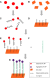
Figure 1
Figure 1
Graphical representation of the biosensor in the detection of Salmonella enterica. Notes: (A) Production of Au NP using the Turkevich method. (B) Synthesis of GO nanosheets through the improved phosphoric acid technique. (C) Primary DNA probe conjugation to Au NP through thiol linkages. (D) Secondary DNA probe conjugation to GO through amide bonding. (E) Both detection platforms are mixed together to form the total biosensor solution. (F) The complementary invA gene target is introduced into the biosensor solution after amplification through polymerase chain reaction. (G) The introduction of the complementary DNA target causes the aggregation of the Au NP-DNA onto the surface of the GO-DNA sheet through hybridization in which the DNA target acts as a cross-linker for both detection platforms. Abbreviations: Au NP, gold nanoparticle; GO, graphene oxide.
Figure 2
Figure 2
Specificity testing of the biosensor utilizing multiple gene targets. Notes: (A) Wavelength analysis of the biosensor solution in comparison with the addition of complementary invA gene target and noncomplementary cspA and myfA gene targets. (B) Visual observation of the biosensor solution color change with the addition of multiple DNA targets.
Figure 3
Figure 3
High-resolution transmission electron microscopy images of the formation of nanocomplex structures between the biosensor detection platforms of Au NP-DNA and GO-DNA with the addition of the invA gene target through DNA hybridization. Notes: (A) Aggregation of Au NP onto a tiny fragmented graphene oxide sheet. (B) Aggregation of Au NP onto a large sheet of graphene oxide. (C) Au NP-GO aggregation visualized at 100 nm magnification (D) Au NP-GO aggregation visualized at 50 nm magnification. Abbreviations: Au NP, gold nanoparticle; GO, graphene oxide.
Figure 4
Figure 4
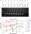
Specificity analysis of the biosensor in elucidating a color change in the detection of various Salmonella enterica sp. Notes: (A) Visual observation of the biosensor color change with the addition of complementary invA gene target from (I) Salmonella typhi, (II) Salmonella covallis, (III) Salmonella heidelberg, (IV) Salmonella typhimurium, (V) Salmonella enteritidis, (VI) Salmonella paratyphi, (VII) Salmonella stanley, (VIII) Salmonella weltevreden, (IX) Salmonella choleraesuis, and (X) without the addition of any DNA target. (B) Gel electrophoresis visualization result on the confirmation of invA gene detection within the Salmonella enterica sp. serovars. (C) Spectrophotometry analysis of the biosensor solution on the detection of multiple serovars of S. enterica.
Figure 5
Figure 5
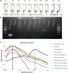
Specificity analysis of the biosensor in elucidating a color change only in the presence of Salmonella enterica sp. with no color change in the presence of other bacterial species. Notes: (A) Visual observation of the biosensor color change with the addition of (I) invA gene target, (II) without the addition of any DNA target material and noncomplementary DNA target from (III) Pseudomonas aeruginosa, (IV) Vibrio cholera, (V) Vibrio parahaemolyticus, (VI) Vibrio proteolyticus, (VII) Staphylococcus aureus, (VIII) Escherichia coli, (IX) Klebsiella pneumoniae, and (X) Streptococcus pneumoniae. (B) Confirmation result of the biosensor specificity analysis through conventional gel electrophoresis method. (C) Spectrophotometry analysis on the biosensor solution with the addition of various bacterial species for specificity analysis on detection.
Figure 6
Figure 6
Sensitivity analysis of the biosensor in elucidating a color change with the minimal amount of target gene required. Notes: (A) Visual observation of the color changing ability by the biosensor solution with the addition of differing concentrations of invA gene target of (I) 0.5 μM, (II) 0.25 μM, (III) 0.125 μM, (IV) 62.5 nM, (V) 31.25 nM, (VI) 15.62 nM, (VII) 7.81 nM, (VIII) 3.91 nM, (IX) 1.96 nM, (X) 0.98 nM, and (XI) 0.49 nM. (B) Spectrophotometry analysis of the biosensor solution with varying concentration levels of DNA target for sensitivity determination.

References

    1. Sheng X, Zhang H, Xia Q, Xu S, Xu H, Huang X. Mig-14 plays an important role in influencing gene expression of Salmonella enterica serovar typhi, which contributes to cell invasion under hyperosmotic conditions. Res Microbiol. 2013;164(9):903–912. - PubMed
    1. Toboldt A, Tietze E, Helmuth R, Junker E, Fruth A, Malorny B. Molecular epidemiology of Salmonella enterica serovar Kottbus isolated in Germany from humans, food and animals. Vet Microbiol. 2014;170(1–2):97–108. - PubMed
    1. Lee C-J, Su L-H, Huang Y-C, Chiu C-H. First isolation of ciprofloxacin-resistant Salmonella enterica serovar typhi in Taiwan. J Microbiol Immunol Infect. 2013;46(6):469–473. - PubMed
    1. Lee SJ, Gebru Awji E, Kim MH, Park SC. BaeR protein from Salmonella enterica serovar paratyphi A induces inflammatory response in murine and human cell lines. Microbes Infect. 2013;15(13):951–957. - PubMed
    1. Crump JA, Mintz ED. Global trends in typhoid and paratyphoid fever. Clin Infect Dis. 2010;50(2):241–246. - PMC - PubMed

Publication types