. 2015 Apr 10:10:171.
doi: 10.1186/s11671-015-0873-8. eCollection 2015.

Application of functional quantum dot nanoparticles as fluorescence probes in cell labeling and tumor diagnostic imaging

Affiliations

Application of functional quantum dot nanoparticles as fluorescence probes in cell labeling and tumor diagnostic imaging

Mei-Xia Zhao et al. Nanoscale Res Lett. .

Abstract

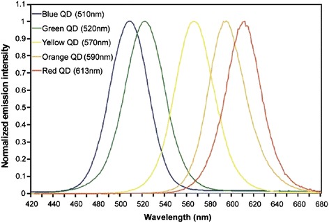
Quantum dots (QDs) are a class of nanomaterials with good optical properties. Compared with organic dyes, QDs have unique photophysical properties: size-tunable light emission, improved signal brightness, resistance against photobleaching, and simultaneous excitation of multiple fluorescence colors. Possessing versatile surface chemistry and superior optical features, QDs are useful in a variety of in vitro and in vivo applications. When linked with targeting biomolecules, QDs can be used to target cell biomarkers because of high luminescence and stability. So QDs have the potential to become a novel class of fluorescent probes. This review outlines the basic properties of QDs, cell fluorescence labeling, and tumor diagnosis imaging and discusses the future directions of QD-focused bionanotechnology research in the life sciences.

Keywords: Biomaterials; Nanoparticles; Optical properties; Quantum dots.


Figures

Figure 1
Figure 1
Various QDs were excited at 350 nm and emission from 420 to 680 nm [ 32 ] .
Figure 2
Figure 2
Fluorescence micrographs of QD-stained cells and tissues. (a) Actin staining on fixed 3 T3 fibroblast, (b) Live MDA-MB-231 breast and (c) mammalian cells. (d) Frozen tissue specimens and a nuclear dye [35].
Figure 3
Figure 3
QD probes enable long-term real-time monitoring of erbB1-eGFP receptor by EGF-QDs. (a) CHO cells expressing erbB1-eGFP after addition of 200 pM 6:1 EGF-QDs. (b) CHO cells expressing erbB1-eGFP after addition of 250 pM 30:1 EGF-QDs. Scale bars 5 μm [37].
Figure 4
Figure 4
Generalized labeling of living cells using QDs. Confocal images of (A) HeLa and (B) D. discoideum cells. (C) Three-dimensional confocal projection of HeLa cells transiently transfected with the plasmid pECFP-Endo (encoding an endosome-specific reporter) and labeled with QD orange as above.[32].
Figure 5
Figure 5
The comparison of intracellular QDs fluorescence intensity of wild-type yeast cells and engineered yeast cells [ 54 ] .
Figure 6
Figure 6
The multicolor imaging of fixed human epithelial cells using five different color QDs [ 55 ] .
Figure 7
Figure 7
Using confocal microscopy for imaging β-CD-L-Arg/CdSe/ZnSe QDs in ECV-304 cells. (a) fluorescent images of Hoechst 33258-stained ECV-304 cells, (b) fluorescent images of ECV-304 cells with β-CD-L-Arg/CdSe/ZnSe QDs, (c) an overlay of bright field and fluorescent images of the β-CD-L-Arg/CdSe/ZnSe QDs labeled ECV-304 cells, (d) in the bright field images. [56]
Figure 8
Figure 8
The schematic of folate-receptor targeted γ-CD/FA-functionalized QDs using tumor-targeting FA in the presence of cell-penetrating β-CD [ 57 ] .
Figure 9
Figure 9
A schematic representation of quantum dot based single cell imaging cytometry for the determination of breast cancer subtypes. Biopsied tissues or primary cells from breast tumors were treated with four different quantum dot-biomarker (EGFR1, HER2, ER, and PR) conjugates and excited by UV light. Hyperspectral fluorescence cellular images were detected at 525 nm, 565 nm, 605 nm, and 655 nm with the help of acousto-optic tunable filter (AOTF). The optical spectrum represents increasing wavelength as a function of color i.e. blue to red. BF: bright field [59].
Figure 10
Figure 10
QD-GA complex-targeted hepatic cells and induced apoptosis through an ROS-mediated mitochondrial dysfunction pathway [ 60 ] .

References

    1. Yong K-T, Law W-C, Hu R, Ye L, Liu L, Swihart MT, et al. Nanotoxicity assessment of quantum dots: from cellular to primate studies. Chem Soc Rev. 2013;42:1236–50. doi: 10.1039/C2CS35392J. - DOI - PubMed
    1. Park Y, Ryu YM, Jung Y, Wang T, Baek Y, Yoon Y, et al. Spraying quantum dot conjugates in the colon of live animals enabled rapid and multiplex cancer diagnosis using endoscopy. ACS Nano. 2014;8:8896–910. doi: 10.1021/nn5009269. - DOI - PubMed
    1. Biju V. Chemical modifications and bioconjugate reactions of nanomaterials for sensing, imaging, drug delivery and therapy. Chem Soc Rev. 2014;43:744–64. doi: 10.1039/C3CS60273G. - DOI - PubMed
    1. Taniguchi S, Green M, Rizvi SB, Seifalian A. The one-pot synthesis of core/shell/shell CdTe/CdSe/ZnSe quantum dots in aqueous media for in vivo deep tissue imaging. J Mater Chem. 2011;21:2877–82. doi: 10.1039/c0jm03527k. - DOI
    1. Chakraborty A, Jana NR. Design and synthesis of triphenylphosphonium functionalized nanoparticle probe for mitochondria targeting and imaging. J Phys Chem C. 2015;119:2888–95.