

Automated characterisation of ultrasound images of ovarian tumours: the diagnostic accuracy of a support vector machine and image processing with a local binary pattern operator
- PMID: 25897367
- PMCID: PMC4402446
Automated characterisation of ultrasound images of ovarian tumours: the diagnostic accuracy of a support vector machine and image processing with a local binary pattern operator
Abstract
Introduction: Preoperative characterisation of ovarian masses into benign or malignant is of paramount importance to optimise patient management.
Objectives: In this study, we developed and validated a computerised model to characterise ovarian masses as benign or malignant.
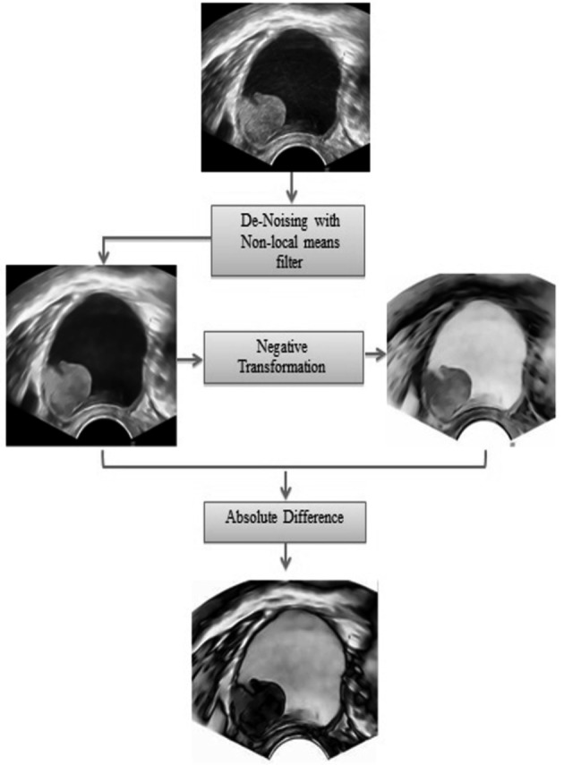
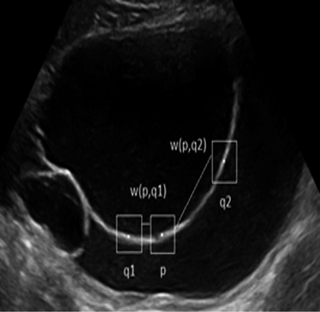
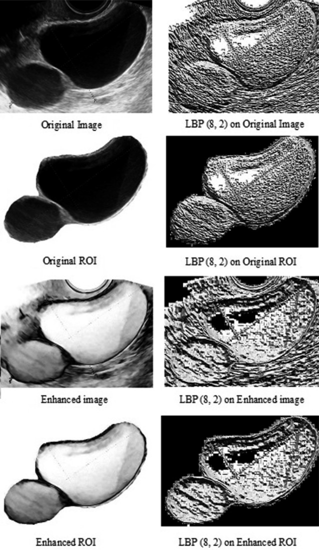
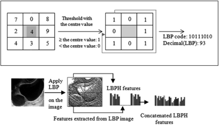
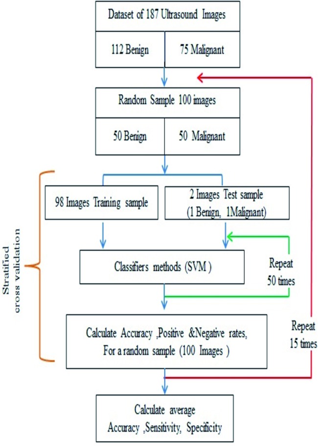
Materials and methods: Transvaginal 2D B mode static ultrasound images of 187 ovarian masses with known histological diagnosis were included. Images were first pre-processed and enhanced, and Local Binary Pattern Histograms were then extracted from 2 × 2 blocks of each image. A Support Vector Machine (SVM) was trained using stratified cross validation with randomised sampling. The process was repeated 15 times and in each round 100 images were randomly selected.
Results: The SVM classified the original non-treated static images as benign or malignant masses with an average accuracy of 0.62 (95% CI: 0.59-0.65). This performance significantly improved to an average accuracy of 0.77 (95% CI: 0.75-0.79) when images were pre-processed, enhanced and treated with a Local Binary Pattern operator (mean difference 0.15: 95% 0.11-0.19, p < 0.0001, two-tailed t test).
Conclusion: We have shown that an SVM can classify static 2D B mode ultrasound images of ovarian masses into benign and malignant categories. The accuracy improves if texture related LBP features extracted from the images are considered.
Keywords: Decision support techniques; Support Vector Machines; ovarian cancer; ovarian neoplasm; ultrasonography.
Figures
References
-
- Acharya UR, Mookiah MR, Vinitha Sree S, et al. Evolutionary algorithm-based classifier parameter tuning for automatic ovarian cancer tissue characterization and classification. Ultraschall Med. 2014;35:237–245. - PubMed
-
- Acharya U, Vinitha Sree S, Saba L, et al. Ovarian tumor characterization and classification: A class of GyneScanTM Systems. Conference proceedings. IEEE Eng Med Biol Soc (EMBC) 2012:4446–4449. - PubMed
-
- Buades A, Coll B, Morel J. A non-local algorithm for image denoising. IEEE Comput Soc Conf Comput Vis Pattern Recognit. 2005;2:60–65.
-
- Biagiotti R, Desii C, Vanzi E, et al. Predicting ovarian malignancy: application of artificial neural networks to transvaginal and color Doppler flow US. Radiology. 1999;210:399–403. - PubMed
-
- Bossuyt PM, Reitsma JB, Bruns DE, et al. Towards complete and accurate reporting of studies of diagnostic accuracy: The STARD Initiative. Radiology. 2003;226:24–28. - PubMed
LinkOut - more resources
Full Text Sources
Miscellaneous