. 2015 Apr 17:7:7.
doi: 10.1186/s13102-015-0002-0. eCollection 2015.

An experimental study of muscular injury repair in a mouse model of notexin-induced lesion with EPI® technique

Affiliations

An experimental study of muscular injury repair in a mouse model of notexin-induced lesion with EPI® technique

Ferran Abat et al. BMC Sports Sci Med Rehabil. .

Abstract

Background: The mechanisms of muscle injury repair after EPI® technique, a treatment based on electrical stimulation, have not been described. This study determines whether EPI® therapy could improve muscle damage.

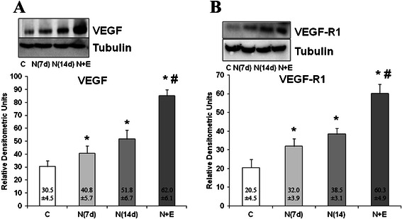
Methods: Twenty-four rats were divided into a control group, Notexin group (7 and 14 days) and a Notexin + EPI group. To induce muscle injury, Notexin was injected in the quadriceps of the left extremity of rats. Pro-inflammatory interleukin 1-beta (IL-1beta) and tumoral necrosis factor-alpha (TNF-alpha) were determined by ELISA. The expression of receptor peroxisome gamma proliferator activator (PPAR-gamma), vascular endothelial growth factor (VEGF) and vascular endothelial growth factor receptor-1 (VEGF-R1) were determined by western-blot.

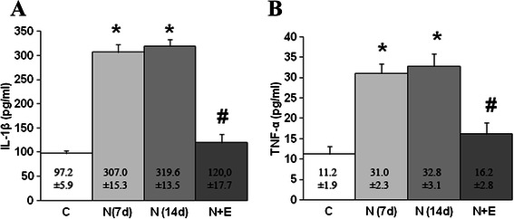
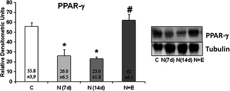
Results: The plasma levels of TNF-alpha and IL-1beta in Notexin-injured rats showed a significant increase compared with the control group. EPI® produced a return of TNF-alpha and IL-1beta values to control levels. PPAR-gamma expression diminished injured quadriceps muscle in rats. EPI® increased PPAR-gamma, VEGF and VEGF-R1 expressions. EPI® decreased plasma levels of pro-inflammatory TNF-alpha and IL-1beta and increased anti-inflammatory PPAR-gamma and proangiogenic factors as well as VEGF and VEGF-R1 expressions.

Conclusion: The EPI® technique may affect inflammatory mediators in damaged muscle tissue and influences the new vascularization of the injured area. These results suggest that EPI® might represent a useful new therapy for the treatment of muscle injuries. Although our study in rats may represent a valid approach to evaluate EPI® treatment, studies designed to determine how the EPI® treatment may affect recovery of injury in humans are needed.

Keywords: EPI; Injury; Muscle; Notexin-induced; Technique.


Figures

Figure 1
Figure 1
Comparison control tissue (A), muscle tissue 21 days after injury induction with Notexina (B) and the effect of the application of EPI® from7 days of the induced lesion (C) in ultrasound imaging (US). It is possible to observe the area of disruption in the middle portion of the quadriceps muscle of rats from 21 days of the induced lesion (surrounded area), compared to normal tissue from the same area (B). Image (C) shows an area of less disruption in the same muscle portion treated with EPI® from 21 days after induction of injury (surrounded area).
Figure 2
Figure 2
Longitudinal ultrasound images of left rat quadriceps. After 7 days treated with Notexin (A), an anechoic image with fluid collection (arrow) indicating muscle lesion was observed. After EPI® treatment (B) a complete resorption of the haematoma with muscle repair (arrow) can be seen.
Figure 3
Figure 3
Plasma levels of IL-1β (A) and TNF-α (B) in control (C), Notexin (N7d, N14 d) and Notexin + EPI (N + E) groups. Values were measured by ELISA assay as indicated in methods. Data are mean ± SD of six independent experiments. *p < 0.05 vs control group; # p < 0.05 vs both Notexin groups.
Figure 4
Figure 4
PPAR-γ protein expression (relative densitometric units) in control (C), Notexin (N7d, N14 d) and Notexin + EPI (N + E) groups. Values were determined in left rat quadriceps muscles by Western blot. A representative inmunoblot is shown and tubulin was used as control amount of protein. Data are mean ± SD of six independent experiments. *p < 0.05 vs control group; # p < 0.05 vs both Notexin groups.
Figure 5
Figure 5
Analysis of VEGF and VEGF-R1 proteins. VEGF (A) and VEGF-R1 (B) protein expression in control (C), Notexin (N7d, N14 d) and Notexin + EPI (N + E) groups were determined by Western blot. Values were determined in left rat quadriceps muscles. In each panel, a representative inmunoblot is shown and tubulin was used as control amount of protein. Data are mean ± SD of six independent experiments. *p < 0.05 vs control group; # p < 0.05 vs both Notexin groups.

References

    1. Ekstrand J, Hägglund M, Waldén M. Epidemiology of muscle injuries in professional football (soccer) Am J Sports Med. 2011;39(6):1226–32. doi: 10.1177/0363546510395879. - DOI - PubMed
    1. Verrall GM, Slavotinek JP, Barnes PG. The effect of sports specific training on reducing the incidence of hamstring injuries in professional Australian rules football players. Br J Sports Med. 2005;39(6):363–8. doi: 10.1136/bjsm.2005.018697. - DOI - PMC - PubMed
    1. Li Y, Foster W, Deasy BM, Chan Y, Prisk V, Tang Y, et al. Transforming growth factor-beta1 induces the differentiation of myogenic cells into fibrotic cells in injured skeletal muscle: a key event in muscle fibrogenesis. Am J Pathol. 2004;164(3):1007–19. doi: 10.1016/S0002-9440(10)63188-4. - DOI - PMC - PubMed
    1. Tidball JG. Inflammatory processes in muscle injury and repair. Am J Physiol Regul Integr Comp Physiol. 2005;288(2):R345–53. doi: 10.1152/ajpregu.00454.2004. - DOI - PubMed
    1. Moresi V, Pristerà A, Scicchitano BM, Molinaro M, Teodori L, Sassoon D, et al. Tumor necrosis factor-alpha inhibition of skeletal muscle regeneration is mediated by a caspase-dependent stem cell response. Stem Cells. 2008;26(4):997–1008. doi: 10.1634/stemcells.2007-0493. - DOI - PubMed

LinkOut - more resources