HER2 and uPAR cooperativity contribute to metastatic phenotype of HER2-positive breast cancer
- PMID: 25897424
- PMCID: PMC4394126
- DOI: 10.18632/oncoscience.146
HER2 and uPAR cooperativity contribute to metastatic phenotype of HER2-positive breast cancer
Abstract
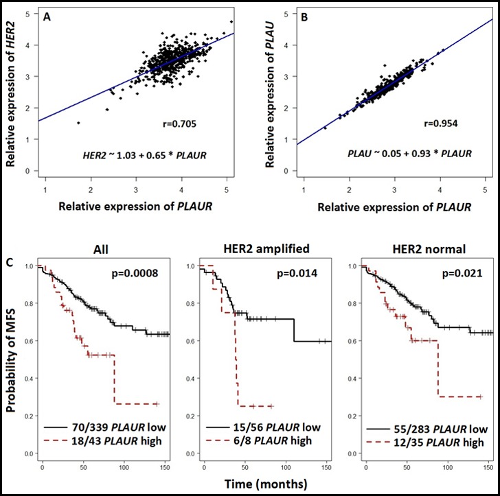
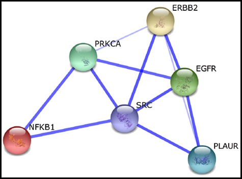
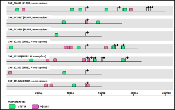
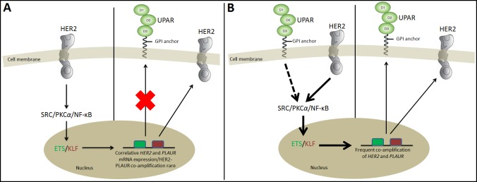
Human epidermal growth factor receptor type 2 (HER2)-positive breast carcinoma is highly aggressive and mostly metastatic in nature though curable/manageable in part by molecular targeted therapy. Recent evidence suggests a subtype of cells within HER2-positive breast tumors that concomitantly expresses the urokinase plasminogen activator receptor (uPAR) with inherent stem cell/mesenchymal-like properties promoting tumor cell motility and a metastatic phenotype. This HER-positive/uPAR-positive subtype may be partially responsible for the failure of HER2-targeted treatment strategies. Herein we discuss and substantiate the cumulative preclinical and clinical evidence on HER2-uPAR cooperativity in terms of gene co-amplification and/or mRNA/protein co-overexpression. We then propose a regulatory signaling model that we hypothesize to maintain upregulation and cooperativity between HER2 and uPAR in aggressive breast cancer. An improved understanding of the HER2/uPAR interaction in breast cancer will provide critical biomolecular information that may help better predict disease course and response to therapy.
Keywords: HER2-positive breast cancer; HER2/ERBB2; co-amplification; co-overexpression; correlation; uPAR/PLAUR.
Conflict of interest statement
No conflict of interests was disclosed by the authors.
Figures
Similar articles
-
RNAi-mediated downregulation of uPAR synergizes with targeting of HER2 through the ERK pathway in breast cancer cells.Int J Cancer. 2010 Oct 1;127(7):1507-16. doi: 10.1002/ijc.25159. Int J Cancer. 2010. PMID: 20063318
-
Combined overexpression of urokinase, urokinase receptor, and plasminogen activator inhibitor-1 is associated with breast cancer progression: an immunohistochemical comparison of normal, benign, and malignant breast tissues.Cancer. 1996 Mar 15;77(6):1079-88. doi: 10.1002/(sici)1097-0142(19960315)77:6<1079::aid-cncr12>3.0.co;2-z. Cancer. 1996. PMID: 8635127
-
uPAR enhances malignant potential of triple-negative breast cancer by directly interacting with uPA and IGF1R.BMC Cancer. 2016 Aug 8;16:615. doi: 10.1186/s12885-016-2663-9. BMC Cancer. 2016. PMID: 27502396 Free PMC article.
-
The d16HER2 Splice Variant: A Friend or Foe of HER2-Positive Cancers?Cancers (Basel). 2019 Jun 28;11(7):902. doi: 10.3390/cancers11070902. Cancers (Basel). 2019. PMID: 31261614 Free PMC article. Review.
-
uPAR: An Essential Factor for Tumor Development.J Cancer. 2021 Oct 17;12(23):7026-7040. doi: 10.7150/jca.62281. eCollection 2021. J Cancer. 2021. PMID: 34729105 Free PMC article. Review.
Cited by
-
N-Alkylisatin-Loaded Liposomes Target the Urokinase Plasminogen Activator System in Breast Cancer.Pharmaceutics. 2020 Jul 7;12(7):641. doi: 10.3390/pharmaceutics12070641. Pharmaceutics. 2020. PMID: 32645963 Free PMC article.
-
High-throughput optofluidic screening of single B cells identifies novel cross-reactive antibodies as inhibitors of uPAR with antibody-dependent effector functions.MAbs. 2023 Jan-Dec;15(1):2184197. doi: 10.1080/19420862.2023.2184197. MAbs. 2023. PMID: 36859773 Free PMC article.
-
Synergistic inhibitory effects of an engineered antibody-like molecule ATF-Fc and trastuzumab on tumor growth and invasion in a human breast cancer xenograft mouse model.Oncol Lett. 2017 Nov;14(5):5189-5196. doi: 10.3892/ol.2017.6896. Epub 2017 Sep 6. Oncol Lett. 2017. PMID: 29113154 Free PMC article.
-
Urokinase-Type Plasminogen Activator Receptor (uPAR) in Inflammation and Disease: A Unique Inflammatory Pathway Activator.Biomedicines. 2024 May 24;12(6):1167. doi: 10.3390/biomedicines12061167. Biomedicines. 2024. PMID: 38927374 Free PMC article. Review.
-
Identification of PLAUR-related ceRNA and immune prognostic signature for kidney renal clear cell carcinoma.Front Oncol. 2022 Aug 16;12:834524. doi: 10.3389/fonc.2022.834524. eCollection 2022. Front Oncol. 2022. PMID: 36052236 Free PMC article.
References
-
- Heiser LM, Sadanandam A, Kuo W-L, Benz SC, Goldstein TC, Ng S, Gibb WJ, Wang NJ, Ziyad S, Tong F, Bayani N, Hu Z, Billig JI, et al. Subtype and pathway specific responses to anticancer compounds in breast cancer. Proceedings of the National Academy of Sciences of the United States of America. 2012;109:2724–9. - PMC - PubMed
-
- Johnston SR. The role of chemotherapy and targeted agents in patients with metastatic breast cancer. European Journal of Cancer. 2011;47:S38–S47. - PubMed
-
- Weigelt B, Peterse JL, van ‘t Veer LJ. Breast cancer metastasis: markers and models. Nature Reviews Cancer. 2005;5:591–602. - PubMed
-
- Harris L, Fritsche H, Mennel R, Norton L, Ravdin P, Taube S, Somerfield MR, Hayes DF, Bast RCJ. American Society of Clinical Oncology update of recommendations for the use of tumor markers in breast cancer. Journal of Clinical Oncology. 2007;25:5287–5312. - PubMed
Publication types
LinkOut - more resources
Full Text Sources
Other Literature Sources
Research Materials
Miscellaneous