. 2015 Feb 9;2(3):272-84.
doi: 10.18632/oncoscience.125. eCollection 2015.

Synergistic enhancement of 5-fluorouracil cytotoxicity by deoxyuridine analogs in cancer cells

Affiliations

Synergistic enhancement of 5-fluorouracil cytotoxicity by deoxyuridine analogs in cancer cells

Yoshihiro Matsumoto et al. Oncoscience. .

Abstract

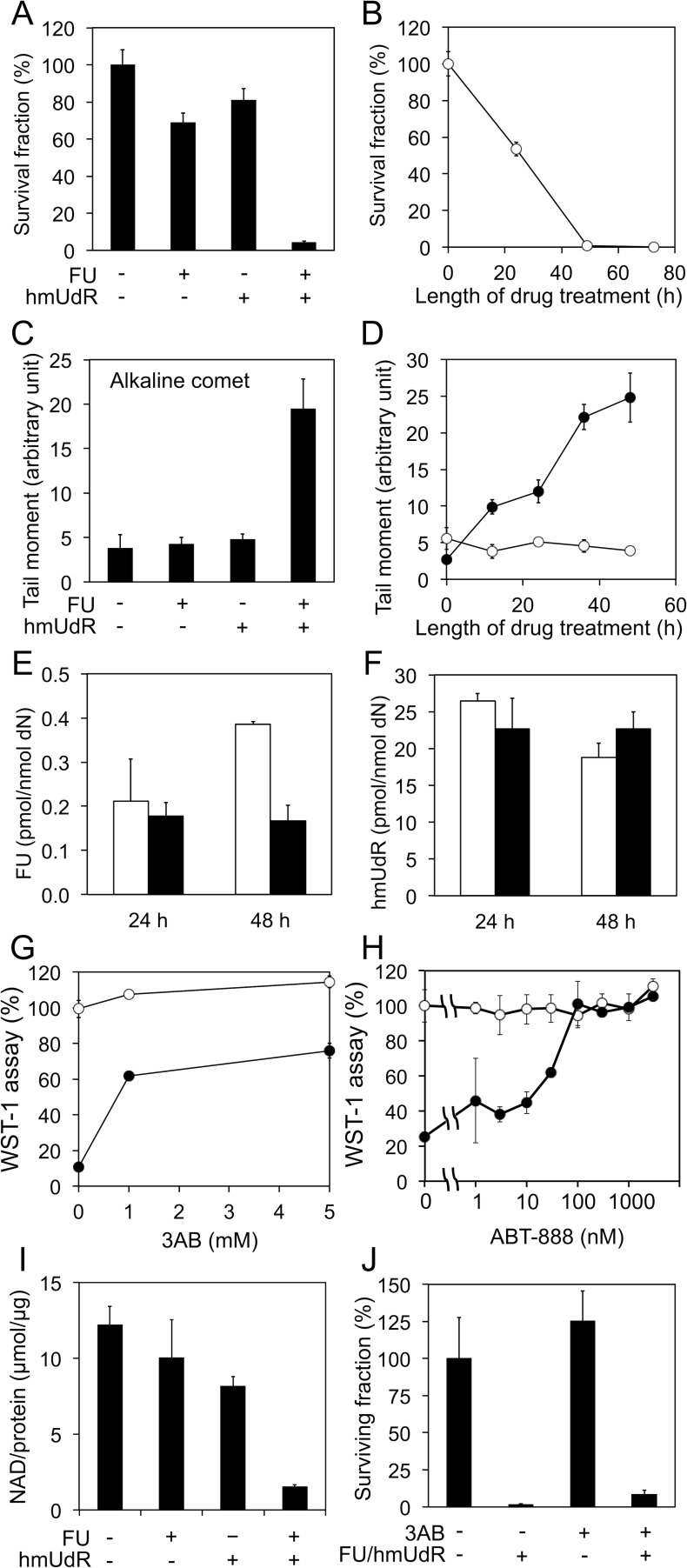
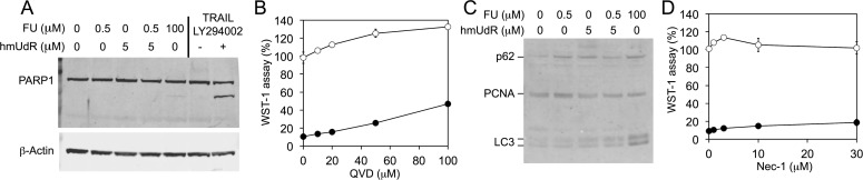
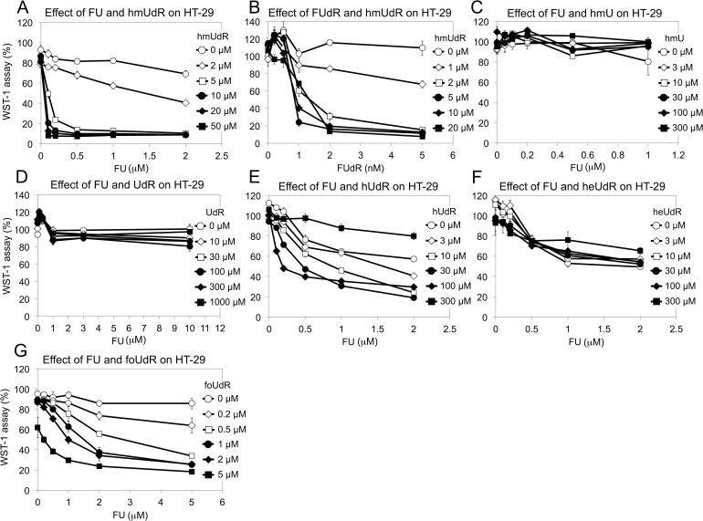
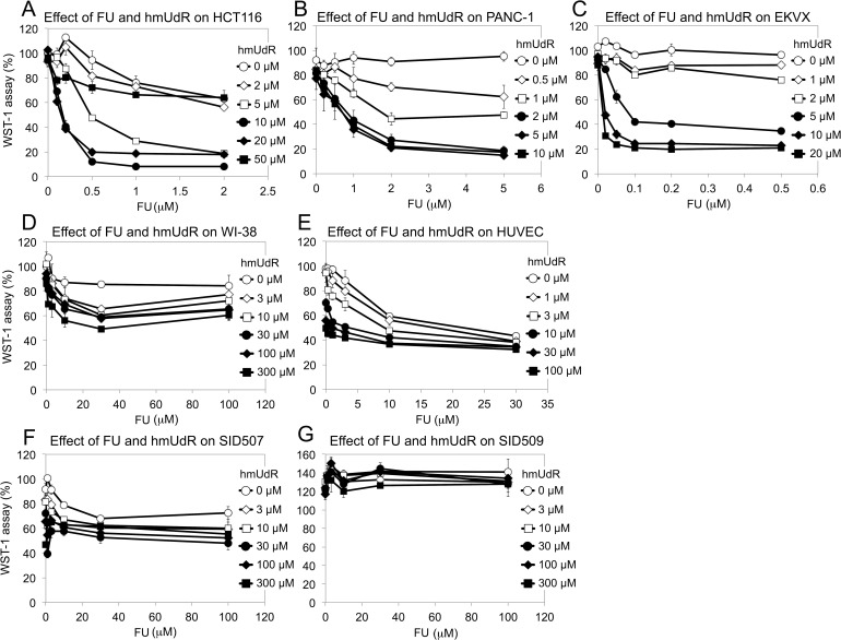
5-Fluorouracil (FU) is a halogenated nucleobase analog that is widely used in chemotherapy. Here we show that 5-hydroxymethyl-2'-deoxyuridine (hmUdR) synergistically enhances the activity of FU in cell lines derived from solid tumors but not normal tissues. While the cytotoxicity of FU and hmUdR was not directly related to the amount of the modified bases incorporated into cellular DNA, incubation with this combination resulted in dramatic increase in the number of single strand breaks in replicating cancer cells, leading to NAD-depletion as consequence of poly(ADP-ribose) synthesis and S phase arrest. Cell death resulting from the base/nucleoside combination did not occur by apoptosis, autophagy or necroptosis. Instead, the cells die via necrosis as a result of NAD depletion. The FU-related nucleoside analog, 5-fluoro-2'-deoxyuridine, also displayed synergy with hmUdR, whereas hmUdR could not be replaced by 5-hydroxymethyluracil. Among other 5-modified deoxyuridine analogs tested, 5-formyl-2'-deoxyuridine and, to a lesser extent, 5-hydroxy-2'-deoxyuridine, also acted synergistically with FU, whereas 5-hydroxyethyl-2'-deoxyuridine did not. Together, our results have revealed an unexpected synergistic interaction between deoxyuridine analogs and FU in a cancer cell-specific manner, and suggest that these novel base/nucleoside combinations could be developed into improved FU-based chemotherapies.

Keywords: 5-Fluorouracil; Cytotoxicity; Deoxyuridine Analog; Synergy.


Conflict of interest statement

CONFLICT OF INTEREST

Two patent applications related to this work were submitted: One by Yoshihiro Matsumoto as an inventor; another by Yoshihiro Matsumoto, Alan E. Tomkinson and Hiroshi Ide as co-inventors. There is no other conflict of interest related to this work.

Figures

Figure 1
Figure 1. Properties of the synergistic toxicity by FU and hmUdR
(A) Colony formation assays of HT-29 cells treated for 48 h with or without 0.5 μM FU and/or 5 μM hmUdR. (B) Time course of effects of FU and hmUdR in colony formation assay. (C) Alkaline comet assays for detection of single-strand breaks (SSBs) in HT-29 cells treated for 48 h with indicated combinations of 0.5 μM FU and 5 μM hmUdR. (D) Time course of SSB formation. The SSB formation was quantitated in HT-29 cells treated with (•) or without (○) 0.5 μM FU and 5 μM hmUdR. (E) Incorporation of FU into HT-29 cellular DNA. Incorporation of tritium-labeled FU (0.5 μM in the medium) was measured in the absence (□) or the presence (■) of 5 μM hmUdR and presented as picomoles per nanomoles of deoxynucleosides. (F) Incorporation of hmUdR into HT-29 cellular DNA. Incorporation of tritium-labeled hmUdR (5 μM in the medium) was measured in the absence (□) or the presence (■) of 0.5 μM FU and presented as picomoles per nanomoles of deoxynucleosides. (G) Effects of 3-aminobenzamide (3AB), a broad PARP inhibitor on the cytotoxicity by FU and hmUdR. 3AB was titrated for its effect on the HT-29 cell growth in the absence (○) or the presence (•) of 0.5 μM FU and 5 μM hmUdR. 3AB was added to the medium simultaneously with FU and hmUdR. The cell growth was measured by WST-1 assay. (H) Effects of ABT-888, a specific inhibitor for PARP1 and PARP2, on the cytotoxicity by FU and hmUdR. ABT-888 was titrated for its effect on the HT-29 cell growth in the absence (○) or the presence (•) of 1 μM FU and 10 μM hmUdR. ABT-888 was added to the medium simultaneously with FU and hmUdR. The cell growth was measured by WST-1 assay. (I) Effect of FU and hmUdR on cellular NAD levels. The quantities of NAD in cell extracts were normalized with the protein concentrations of the extracts. (J) Survival fractions of HT-29 cells treated with drugs in the presence of 3AB for 72 h. After replating without drugs, the cells were allowed to grow for 6 days and their nucleic acids were quantitated by CyQUANT kit. Data in panels A-J are from triplicate experiments and plotted with standard deviations.
Figure 2
Figure 2. Cell cycle analyses of HT-29 cells by flow cytometry
(A) Time course of cell cycle distribution of synchronized cells treated with a combination of 0.5 μM FU and 5 μM hmUdR. HT-29 cells were synchronized at the G /S boundary by sequential pretreatment with nocodazole and aphidicolin as described in Materials and Methods. The time at which aphidicolin was removed is designated 0 h. When indicated, FU and hmUdR were added through aphidicolin treatment and subsequent incubation. (B) Effect of FU, hmUdR and caffeine on cell cycle distribution. Unsynchronized HT-29 cells were treated without or with 0.5 μM FU and 5 μM hmUdR for 48 h, and incubated in the absence or presence of 5 mM caffeine for the last 24 h. (C) Cell cycle analyses of unsynchronized HT-29 cells in the presence of 3AB and caffeine. (D) Alkaline comet assay of HT-29 cells treated for 48 h with drugs in the presence of 3AB. In both experiments, 0.5 μM FU, 5 μM hmUdR and 3 mM 3AB were added when indicated. Data in panel D are from triplicate experiments and plotted with standard deviations.
Figure 3
Figure 3. Characterization of the mechanism for cell death resulting from combined treatment with FU and hmUdR
(A) Immunoblot detection of PARP1. PARP1 cleavage was examined in whole cell extracts of HT-29 cells treated for 72 h with indicated concentrations of FU and hmUdR. As a positive control for PARP1 cleavage, HT-29 cells were treated with 50 μM LY294002 for 1 h followed by 4 h treatment with 100 μg/ml TRAIL. β-Actin was a loading control. (B) Effects of an apoptosis inhibitor. A broad spectrum caspase inhibitor, QVD, were tested for their effects on the HT-29 cell growth in the absence (○) or the presence (•) of 0.5 μM FU and 5 μM hmUdR. QVD was added to the medium simultaneously with FU and hmUdR. The cell growth was measured by WST-1 assay. The slight increase in cell growth with 50 and 100 μM QVD was an effect of DMSO in which QVD was dissolved. (C) Immunoblot detection of autophagy-related proteins, p62 and LC3 (microtubule-associated protein 1 light chain 3). p62, LC3 and a loading control, PCNA, were detected in the whole cell extracts prepared by the same way as for panel A. Autophagy is expected to decrease p62 and increase the LC3 proteins. (D) Effects of a necroptosis inhibitor on the cytotoxicity by FU and hmUdR. Necrostatin-1 (Nec-1) was tested for their effects on the HT-29 cell growth in the absence (○) or the presence (•) of 0.5 μM FU and 5 μM hmUdR. Nec-1 was added to the medium simultaneously with FU and hmUdR. The cell growth was measured by WST-1 assay. Data in panels B and D are from triplicate experiments and plotted with standard deviations.
Figure 4
Figure 4. Chemical structures of base/nucleoside analogs tested in this study
(A) FU. (B) hmUdR. (C) FUdR. (D) hmU. (E) hUdR. (F) heUdR. (G) foUdR.
Figure 5
Figure 5. Effect of various drug combinations on the growth of HT-29 cells
(A) FU and hmUdR. (B) 5-fluoro-2′-deoxyuridine (FUdR) and hmUdR. (C) FU and hmU. (D) FU and 2′-deoxyuridine (UdR). (E) 5-hydroxy-2′-deoxyuridine (hUdR) and FU. (F) 5-hydroxyethyl-2′-deoxyuridine (heUdR) and FU. (G) 5-formyl-2′-deoxyuridine (foUdR) and FU. HT-29 cells were treated with indicated compounds for 72 hours, and the cell proliferations were measured by WST-1 assay. Data are from triplicate experiments and plotted with standard deviations.
Figure 6
Figure 6. Effect of FU and hmUdR on the growth of various cells
(A) HCT 116 (p53-proficient colorectal carcinoma). (B) PANC-1 (pancreatic cancer). (C) EKVX (non-small cell lung cancer). (D) A normal cell line, WI-38 (embryonic lung fibroblast). (E) Human umbilical vein endothelial cells (HUVEC). These cells were treated for 72 hours with increasing concentrations of FU and hmUdR, and their proliferations were measured by WST-1 assay. (F) SID507 (normal human colon cell line). (G) SID509 (normal human colon cell line). These normal colon cells were tested by the same procedures as above except that they were incubated with or without FU and hmUdR for 7 days. Data are from triplicate experiments and plotted with standard deviations.

Similar articles

Cited by

References

    1. Grem JL. 5-Fluorouracil: forty-plus and still ticking. A review of its preclinical and clinical development. Investigational New Drugs. 2000;18:299–313. - PubMed
    1. O'Connor OA. Pharmacological Modulation of Fluoropyrimidines: Building on the Lessons of the Past. In: Schwartz GK, editor. Combination Cancer Therapy: Modulators and Potentiators. Totowa, NJ: Humana Press; 2005. pp. 133–174.
    1. Yoshida K, Yamaguchi K, Osada S, Kawaguchi Y, Takahashi T, Sakashita F, Tanaka Y. Challenge for a better combination with basic evidence. Int J Clin Oncol. 2008;13:212–219. - PubMed
    1. Lewis HL, Muhleman DR, Ward JF. Serologic assay of DNA base damage. I. 5-Hydroxymethyldeoxyuridine, a radiation product of thymidine. Radiation Research. 1978;75:305–316. - PubMed
    1. Teebor GW, Frenkel K, Goldstein MS. Ionizing radiation and tritium transmutation both cause formation of 5-Hydroxymethyl-2′-deoxyuridine in cellular DNA. Proc Natl Acad Sci USA. 1984;81:318–321. - PMC - PubMed

LinkOut - more resources