. 2015 Mar 23;2(3):309-16.
doi: 10.18632/oncoscience.144. eCollection 2015.

Bone morphogenetic protein 4 (BMP-4) and epidermal growth factor (EGF) inhibit metalloproteinase-9 (MMP-9) expression in cancer cells

Affiliations

Bone morphogenetic protein 4 (BMP-4) and epidermal growth factor (EGF) inhibit metalloproteinase-9 (MMP-9) expression in cancer cells

Nathalie Bibens Laulan et al. Oncoscience. .

Abstract

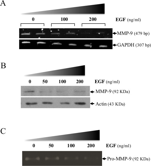
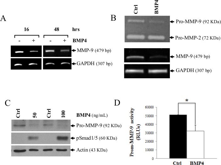
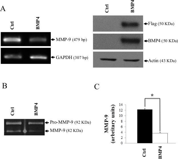
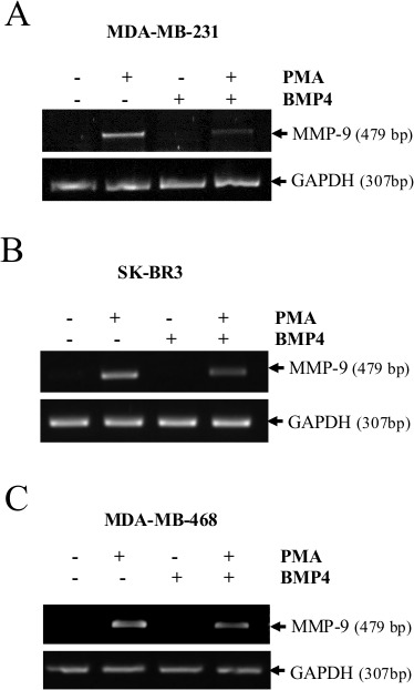
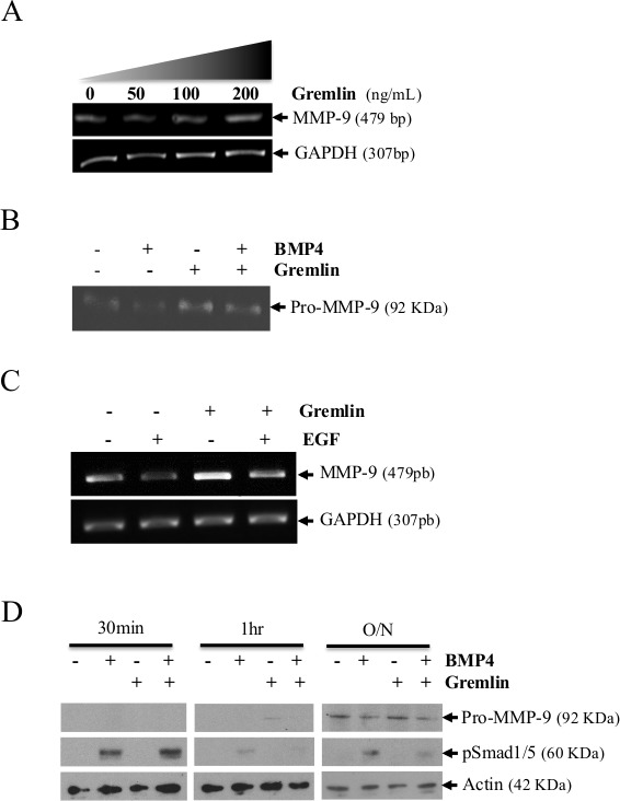
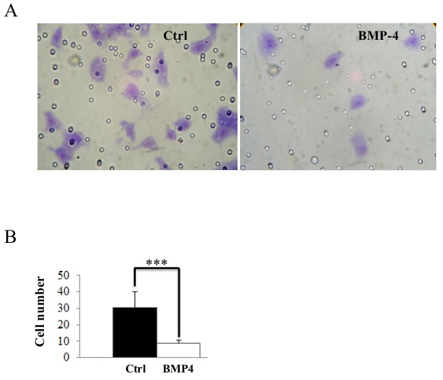
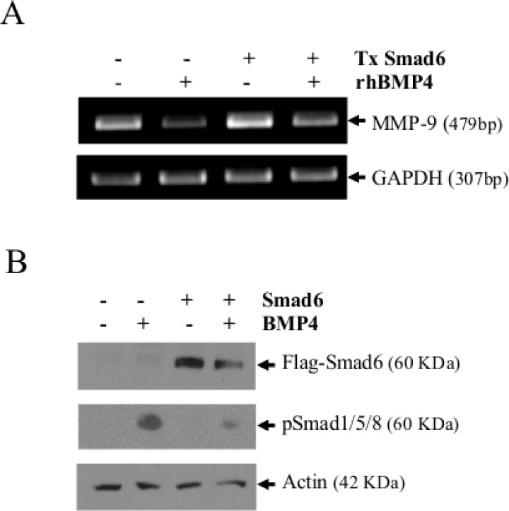
Matrix metalloproteinase-9 (MMP-9) plays a central role in the progression of the cancer. While a large number of studies have contributed to our understanding of the molecular mechanisms responsible for upregulating MMP-9 gene expression in normal and cancer cells, our knowledge on the signals that suppress MMP-9 expression is much more limited. Here, we report that EGF and BMP-4 cooperate to inhibit MMP-9 expression in cancer cells. Treatment with EGF reduces the expression of MMP-9 at both mRNA while augmenting BMP-4 expression. Interestingly, recombinant BMP-4 suppressed constitutive and PMA-induced MMP-9 expression in both fibrosarcoma and breast cancer cells. Addition of gremlin a natural inhibitor of BMP-4, inhibited the suppression of MMP-9 by EGF. The suppression of MMP-9 by BMP-4 likely occurs at the transcriptional level since BMP-4 suppressed MMP-9 mRNA expression and activation of a reporter vector encoding the human MMP-9 promoter. The suppressive effect of BMP-4 occurs via Smad1/5/8 and is specific since BMP-4 did not inhibit MMP-2 while BMP-2 was ineffective in suppressing MMP-9. Taken together, these results are consistent with a new paradigm for the role of EGF and BMPs in controlling MMP gene expression in cancer cells.


Conflict of interest statement

CONFLICT OF INTEREST

The authors of this manuscript have no conflicts of interest to declare.

Figures

Figure 1
Figure 1. Suppression of MMP-9 in HT1080 cells following treatment with EGF
(A) MMP-9 mRNA expression in absence or presence of recombinant EGF. Levels of transcripts were measured 16 h after adding EGF. GAPDH was used as loading and specificity control. Suppression of MMP-9 expression by increasing doses of EGF was confirmed at the protein level using (B) Western-blot analysis and (C) gelatin zymography. Data are representative of at least two independent experiments.
Figure 2
Figure 2. BMP-4 expression in HT1080 cells by EGF/EGR1
(A) BMP-4 mRNA expression in absence or presence of recombinant EGF. Levels of transcripts were measured 16 h after adding EGF. GAPDH was used as loading and specificity control. (B), BMP-4 and MMP-9 expression in HT1080 cells following stable expression of an expression vector encoding human EGR1. GAPDH was used as loading and specificity control. Data are representative of at least two independent experiments.
Figure 3
Figure 3. MMP-9 is decreased following BMP-4 stimulation in HT1080 cells
(A) MMP-9 mRNA expression 24 and 48h following stimulation with recombinant BMP-4 (200 ng/ml). GAPDH was used as loading and specificity control. In (B), a zymogram (top gel) showing reduced MMP-9 secretion in supernatants of HT1080 cells treated for 16h with recombinant BMP-4 (200ng/ml). The lower panel shows the MMP-9 mRNA level of the treated cells. (C) Western blot analysis showing expression of MMP-9 and phosphorylation of Smad1/5 after treatment with human recombinant BMP-4. (D) Luciferase activity of in HT1080 cells transfected with a luciferase reporter vector containing the MMP-9-promoter following treatment with human recombinant BMP-4. Statistical analyses were carried out using Student's t test for unpaired samples. (* = p ≤ 0,05; ** = p ≤ 0,005).
Figure 4
Figure 4. De novo expression of BMP-4 reduces MMP-9 gene expression
(A) MMP-9 mRNA expression in mock-transfected HT1080 cells or HT1080 cells transfected with an expression vector encoding a flagged human BMP-4 (pCMV-BMP-4-Flag). GAPDH was used as loading and specificity control. The lower panel represents the control Western blot gels showing de novo expression of flagged BMP-4 in transfected cells. (B) Zymography (left panel) showing MMP-9 in the supernatant of transfected cells as in (A). (C) Quantitative analyses of MMP-9 expression by imaging densitometry is shown on the right histogram, which represents the means of independent experiments shown in (A). Data are representative of at least three independent experiments. (* = p ≤ 0,05)
Figure 5
Figure 5. MMP-9 expression is suppressed by BMP-4 in MDA-MB-231, MDA-MB-468 and SKBR3 cells
MMP-9 mRNA expression in PMA-stimulated cells (20ng/ml) treated with or without recombinant BMP-4 (50 or 100 ng/ml) in MDA-MB-231 cells (A), SK-BR3 (B) and MDA-MB-468 cells (C). GAPDH was used as loading and specificity control. Data are representative of at least three independent experiments.
Figure 6
Figure 6. MMP-9 is increased following treatment of HT1080 cells with gremlin
(A) MMP-9 mRNA expression in cells treated with increasing concentrations of gremlin. GAPDH was used as loading and specificity control. (B) Zymogram showing MMP-9 in the supernatant of HT1080 cells treated with recombinant BMP-4 in absence or presence of gremlin. (C) Kinetic analysis showing MMP-9 expression and phosphorylation of Smad1/5 in cells treated treatment with human recombinant BMP-4 with or without gremlin. Data are representative of at least three independent experiments. (D) MMP-9 mRNA expression in absence or presence of recombinant EGF and gremlin in HT1080 cells.
Figure 7
Figure 7. BMP-4 inhibits in vitro HT1080 cell invasion
(A) HT1080 cell migration across Matrigel with or without recombinant BMP-4. (B) Migrated HT1080 cells were quantified. Data are average ± SEM; n = 10 unit areas. Data are representative of at least three independent experiments. (***= p ≤ 0,001).
Figure 8
Figure 8. Smad6 reduces the ability of BMP-4 to suppress MMP-9 expression
(A) MMP-9 mRNA expression in mock-transfected HT1080 cells or HT1080 cells transfected with an expression vector encoding a flagged Smad6 (pCS2-Smad6-Flag). MMP-9 expression was measured 16h after transfection. The lower panel represents the control Western blot gels showing de novo expression of Smad6 in transfected cells. GAPDH and actin were used as loading and specificity controls for RT-PCR and Western blot analyses respectively. In (B), a zymogram showing MMP-9 in the supernatant of HT1080 transfected cells. Data are representative of at least three independent experiments.

References

    1. Bernhard EJ, Gruber SB, Muschel RJ. Direct evidence linking expression of matrix metalloproteinase 9 (92-kDa gelatinase/collagenase) to the metastatic phenotype in transformed rat embryo cells. Proc Natl Acad Sci U S A. 1994;91:4293–4297. - PMC - PubMed
    1. Egeblad M, Werb Z. New functions for the matrix metalloproteinases in cancer progression. Nat Rev Cancer. 2002;2:161–174. - PubMed
    1. Kessenbrock K, Plaks V, Werb Z. Matrix metalloproteinases: regulators of the tumor microenvironment. Cell. 2010;141:52–67. - PMC - PubMed
    1. Lopez-Otin C, Matrisian LM. Emerging roles of proteases in tumour suppression. Nat Rev Cancer. 2007;7:800–808. - PubMed
    1. Chicoine E, Esteve PO, Robledo O, Van Themsche C, Potworowski EF, St-Pierre Y. Evidence for the role of promoter methylation in the regulation of MMP-9 gene expression. Biochem Biophys Res Commun. 2002;297:765–772. - PubMed

LinkOut - more resources