. 2015 May;21(5):856-9.
doi: 10.3201/eid2105.150041.

Postmortem stability of Ebola virus

Postmortem stability of Ebola virus

Joseph Prescott et al. Emerg Infect Dis. 2015 May.

Abstract

The ongoing Ebola virus outbreak in West Africa has highlighted questions regarding stability of the virus and detection of RNA from corpses. We used Ebola virus-infected macaques to model humans who died of Ebola virus disease. Viable virus was isolated <7 days posteuthanasia; viral RNA was detectable for 10 weeks.

Keywords: Ebola hemorrhagic fever; Ebola virus; Ebola virus disease; West Africa; cynomolgus macaques; outbreak; postmortem stability; transmission; viruses.


Figures
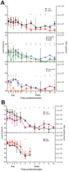
Figure 1
Figure 1
Presence and stability of Ebola virus RNA in deceased cynomolgus macaques. Swab (A) and tissue (B) specimen samples were obtained at the indicated time points, and viral RNA was isolated and used in a 1-step quantitative reverse transcription PCR with a primer/probe set specific for the nucleoprotein gene and standards consisting of known nucleoprotein gene copy numbers. Line plots show means of positive samples from 5 animals up to the 7 day time point and from 3 animals thereafter. Error bars indicate SD, and - indicates time points at which ≥1 animal had undetectable levels of viral RNA. Absence of a hyphen indicates that all animals had detectible levels of viral RNA.
Figure 2
Figure 2
Efficiency of Ebola virus isolation from deceased cynomolgus macaques. Swab (A) and tissue (B) specimen samples were obtained at the indicated time points, and virus isolation was attempted on Vero E6 cells. Cells were inoculated in triplicate with serial dilutions of inoculum from swab specimens placed in 1 mL of medium or tissues homogenized in 1 mL of medium. The 50% tissue culture infectious dose (TCID50) was calculated by using the Spearman-Karber method (8). Line plots show means of positive samples from 5 animals to the day 9 time point. Error bars indicate SD.

References

    1. Centers for Disease Control and Prevention. Ebola hemorrhagic fever [cited 2015 Jan 3]. http://www.cdc.gov.ezproxy.nihlibrary.nih.gov/vhf/ebola/
    1. Dowell SF, Mukunu R, Ksiazek TG, Khan AS, Rollin PE, Peters CJ. Transmission of Ebola hemorrhagic fever: a study of risk factors in family members, Kikwit, Democratic Republic of the Congo, 1995. J Infect Dis. 1999;179(Suppl 1):S87–91 . 10.1086/514284 - DOI - PubMed
    1. Khan AS, Tshioko FK, Heymann DL, Le Guenno B, Nabeth P, Kerstiëns B, et al. The reemergence of Ebola hemorrhagic fever, Democratic Republic of the Congo, 1995. J Infect Dis. 1999;179(Suppl 1):S76–86. 10.1086/514306 - DOI - PubMed
    1. Hoenen T, Groseth A, Feldmann F, Marzi A, Ebihara H, Kobinger G, et al. Complete genome sequences of three Ebola virus isolates from the 2014 outbreak in West Africa. Genome Announc. 2014;2:e01331–14. - PMC - PubMed
    1. Ng S, Cowling B. Association between temperature, humidity and ebolavirus disease outbreaks in Africa, 1976 to 2014. Euro Surveill. 2014;19:20892 . - PubMed

Publication types