. 2015 May 19;54(19):3037-3050.
doi: 10.1021/acs.biochem.5b00150. Epub 2015 May 4.

Kinetic and Structural Basis for Acyl-Group Selectivity and NAD(+) Dependence in Sirtuin-Catalyzed Deacylation

Affiliations

Kinetic and Structural Basis for Acyl-Group Selectivity and NAD(+) Dependence in Sirtuin-Catalyzed Deacylation

Jessica L Feldman et al. Biochemistry. .

Abstract

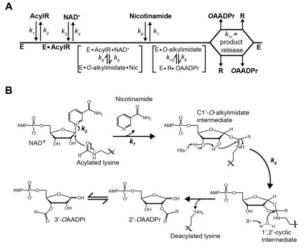
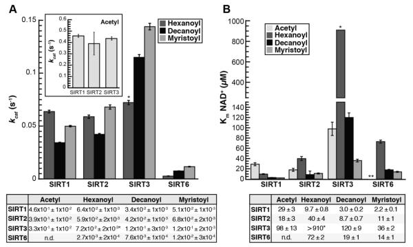
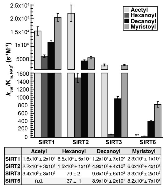
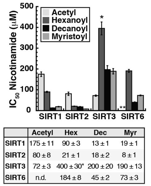
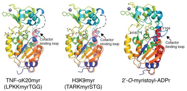
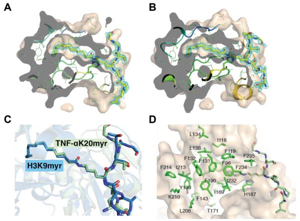
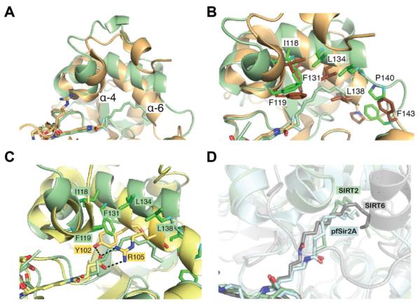
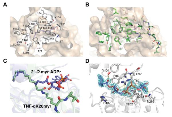
Acylation of lysine is an important protein modification regulating diverse biological processes. It was recently demonstrated that members of the human Sirtuin family are capable of catalyzing long chain deacylation, in addition to the well-known NAD(+)-dependent deacetylation activity [Feldman, J. L., Baeza, J., and Denu, J. M. (2013) J. Biol. Chem. 288, 31350-31356]. Here we provide a detailed kinetic and structural analysis that describes the interdependence of NAD(+)-binding and acyl-group selectivity for a diverse series of human Sirtuins, SIRT1-SIRT3 and SIRT6. Steady-state and rapid-quench kinetic analyses indicated that differences in NAD(+) saturation and susceptibility to nicotinamide inhibition reflect unique kinetic behavior displayed by each Sirtuin and depend on acyl substrate chain length. Though the rate of nucleophilic attack of the 2'-hydroxyl on the C1'-O-alkylimidate intermediate varies with acyl substrate chain length, this step remains rate-determining for SIRT2 and SIRT3; however, for SIRT6, this step is no longer rate-limiting for long chain substrates. Cocrystallization of SIRT2 with myristoylated peptide and NAD(+) yielded a co-complex structure with reaction product 2'-O-myristoyl-ADP-ribose, revealing a latent hydrophobic cavity to accommodate the long chain acyl group, and suggesting a general mechanism for long chain deacylation. Comparing two separately determined co-complex structures containing either a myristoylated peptide or 2'-O-myristoyl-ADP-ribose indicates there are conformational changes at the myristoyl-ribose linkage with minimal structural differences in the enzyme active site. During the deacylation reaction, the fatty acyl group is held in a relatively fixed position. We describe a kinetic and structural model to explain how various Sirtuins display unique acyl substrate preferences and how different reaction kinetics influence NAD(+) dependence. The biological implications are discussed.


Figures

Figure 1
Figure 1. Proposed Sirtuin deacylation mechanism and kinetic scheme
A, Sirtuin enzymes follow a sequential mechanism in which both acylated substrate (AcylR) (k1) and NAD+ (k3) bind prior to any catalytic step. SIRT1, SIRT2 and SIRT3 must bind AcylR prior to NAD+, while SIRT6 is the only mammalian Sirtuin capable of binding AcylR or NAD+ in random order. A ternary complex is formed, followed by nicotinamide formation (k5) and release (k7), and transfer of the acyl group from AcylR to ADP-ribose (k9). Deacylated substrate (R) and O-acyl-ADPr (OAADPr) are randomly released (k11). B, Proposed deacylation mechanism. Nucleophilic addition of the acyl oxygen on the 1′-carbon of the nicotinamide ribose forms the C1′-O-alkylimidate intermediate. A conserved histidine residue in the active site activates the 2′-hydroxyl group of NAD+ ribose. The activated hydroxyl attacks the O-alkylimidate carbon to afford the 1′2′-cyclic intermediate. A base activated water molecule attacks the cyclic intermediate resulting in the formation of deacylated lysine and OAADPr. Rate constants are indicated.
Figure 2
Figure 2. kcat and Km, NAD for SIRT1, SIRT2, SIRT3, and SIRT6 deacylation reactions
A, kcat, inset: kcat for acetylated substrate plotted separately for clarity; Calculated kcat values determined from non-linear regression fits to Michaelis-Menten shown below (n ≥ 3, ± standard deviation). *Estimate for SIRT3 resulting from inability to saturate reaction with NAD+. B, Km for NAD+, Calculated Km, NAD values determined from non-linear regression fits to Michaelis-Menten shown below. *Estimate for SIRT3, **Km, NAD with acetylated peptide for SIRT6 was not measured due to prohibitively high peptide substrate necessary to saturate the reaction (n ≥ 3, ± standard deviation of mean).
Figure 3
Figure 3. kcat/Km,NAD for SIRT1, SIRT2, SIRT3, and SIRT6 deacylation reactions
kcat/Km,NAD determined for SIRT1, SIRT2, SIRT3, and SIRT6 in the presence of acetyl-, hexanoyl-, decanoyl-, and myristoyl-lysine H3K9 peptides. Calculated parameters determined from non-linear regression fits to Michaelis-Menten shown below. **kcat/Km,NAD with acetylated peptide for SIRT6 was not measured due to prohibitively high peptide substrate necessary to saturate the reaction (n ≥ 3, ± standard deviation of mean)
Figure 4
Figure 4. Nicotinamide IC50 values
SIRT1, SIRT2, SIRT3 and SIRT6 display varied susceptibilities to nicotinamide inhibition dependent upon acyl-substrate (n ≥ 2, ± standard deviation). *Due to the high Km for NAD+ in the presence of the hexanoylated peptide (>910 μM) the assay was performed in the presence of 800 μM NAD+. **Nicotinamide IC50 for SIRT6 in the presence of acetylated peptide was not measured due to prohibitively high peptide substrate concentration necessary to saturate the reaction.
Figure 5
Figure 5. Crystal structure of SIRT2 in complex with TNFα-K20myr, H3K9myr or 2′-O-myristoyl-ADP-ribose
Overall structural features of SIRT2 in the presence of myristoylated substrates or product. The cofactor binding loop was not visible in the SIRT2 – TNFα-K20myr or SIRT2 – H3K9myr structures and is shown as a dashed line for clarity. Also highlighted in the 2′-O-myristoyl-ADPr structure are the locations and orientations of Tyr104 and the catalytic histidine, His187.
Figure 6
Figure 6. Analysis of myristoyl-lysine binding pocket
A, 2Fo-Fc omit electron density map (cyan mesh, 1σ) of the TNF-αK20 myristoylated peptide. Molecular surface of SIRT2 cut at the level of the hydrophobic cavity are shown. The myristoylated peptides are drawn as sticks, in which yellow, blue and red represent C, N and O atoms, respectively. B, 2Fo-Fc omit electron density map (cyan mesh, 1σ) of the H3K9 myristoylated peptide represented as shown in A. C, superimposition of SIRT2 – TNFα-K20myr (light green) or SIRT2 – H3K9myr (blue) highlighting the orientation of the myristoylated lysine chain. D, hydrophobic pocket in SIRT2 that accommodates the myristoyl chain.
Figure 7
Figure 7. SIRT2 – TNFα-K20myr structural comparison
A, superimposition of SIRT2 - TNFα-K20myr (light green) with SIRT2 - ε-trifluoroacetyl lysine peptide inhibitor (PDB ID: 4L3O, brown), highlighting the differences in the helix bundle region. B, same structures as in A, highlighting the orientations of specific amino acids. C, superimposition of SIRT2 – TNFα-K20myr (light green) with SIRT5- H3K9succinyl (PDB: 4F4U, yellow). The amino acids in α-helix 6 that interact with the peptide substrates are shown in stick representation. D, superimposition of SIRT2 – TNFα-K20myr (light green) with SIRT6 – H3K9myr (PDB ID: 3ZG6, light blue) and pfSir2A – H3K9myr (PDB ID: 3U3D, grey), highlighting the various orientations of the myristoyl group.
Figure 8
Figure 8. Comparison of SIRT2 – 2′-O-myristoyl-ADPr with SIRT2 – TNFα-K20myr structure
A, hydrophobic pocket in SIRT2 that accommodates the myristoyl chain of 2′-O-myristoyl-ADPr. B, superimposition of SIRT2 –2′-O-myristoyl-ADPr (grey) and SIRT2 – TNFα-K20myr (green) structures highlighting the hydrophobic residues in the myristoyl chain binding site. C, overlay as in B highlighting differences in myristoyl chain between substrate and product. D, 2Fo-Fc omit electron density map (blue mesh, 1σ) of 2′-O-myristoyl-ADPr molecule and hydrogen bonding network surrounding 2′-O-myristoyl-ADPr. Also shown is the catalytic histidine residue.

Similar articles

Cited by

References

    1. Feldman JL, Baeza J, Denu JM. Activation of the protein deacetylase SIRT6 by long-chain fatty acids and widespread deacylation by mammalian sirtuins. J Biol Chem. 2013;288:31350–31356. - PMC - PubMed
    1. Jiang H, Khan S, Wang Y, Charron G, He B, Sebastian C, Du J, Kim R, Ge E, Mostoslavsky R, Hang HC, Hao Q, Lin H. SIRT6 regulates TNF-alpha secretion through hydrolysis of long-chain fatty acyl lysine. Nature. 2013;496:110–113. - PMC - PubMed
    1. Wagner GR, Hirschey MD. Nonenzymatic protein acylation as a carbon stress regulated by sirtuin deacylases. Mol Cell. 2014;54:5–16. - PMC - PubMed
    1. He W, Newman JC, Wang MZ, Ho L, Verdin E. Mitochondrial sirtuins: regulators of protein acylation and metabolism. Trends Endocrinol Metab. 2012;23:467–476. - PubMed
    1. Cai L, Sutter BM, Li B, Tu BP. Acetyl-CoA induces cell growth and proliferation by promoting the acetylation of histones at growth genes. Mol Cell. 2011;42:426–437. - PMC - PubMed

Publication types