. 2015 Apr 21;10(4):e0124296.
doi: 10.1371/journal.pone.0124296. eCollection 2015.

Exposure to endocrine disruptor induces transgenerational epigenetic deregulation of microRNAs in primordial germ cells

Affiliations

Exposure to endocrine disruptor induces transgenerational epigenetic deregulation of microRNAs in primordial germ cells

Miguel A Brieño-Enríquez et al. PLoS One. .

Abstract

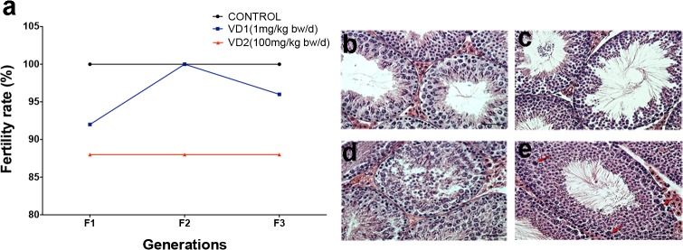
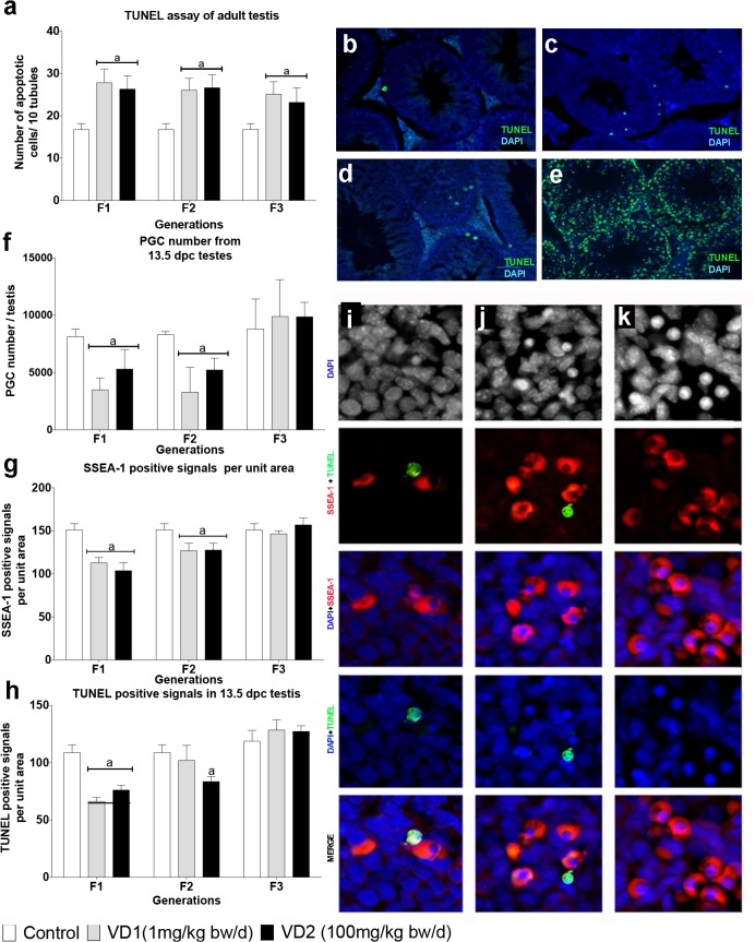
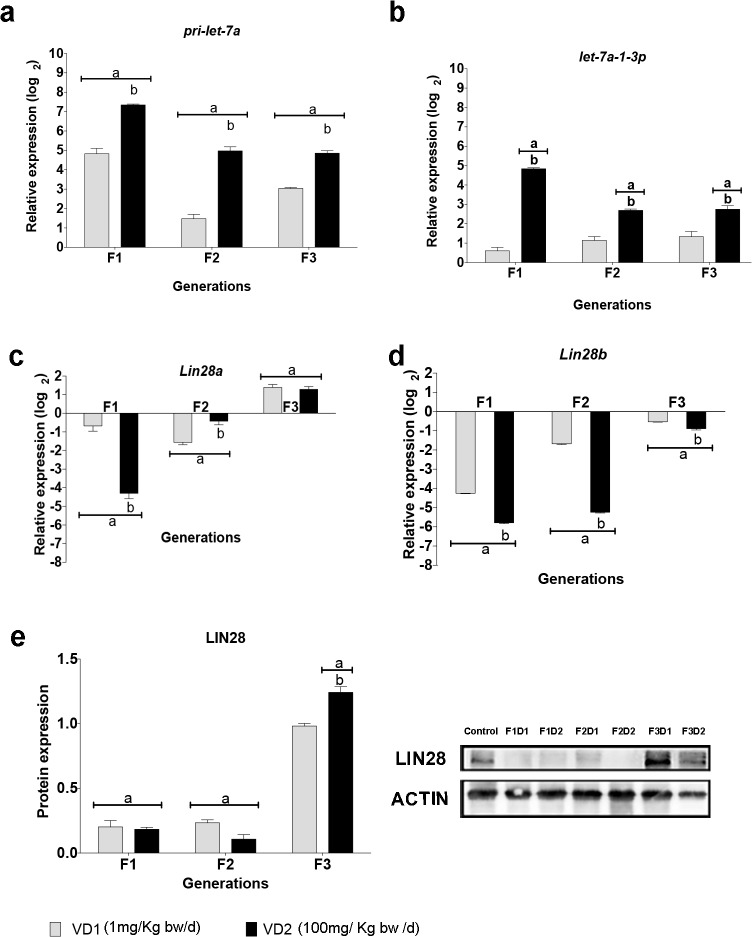
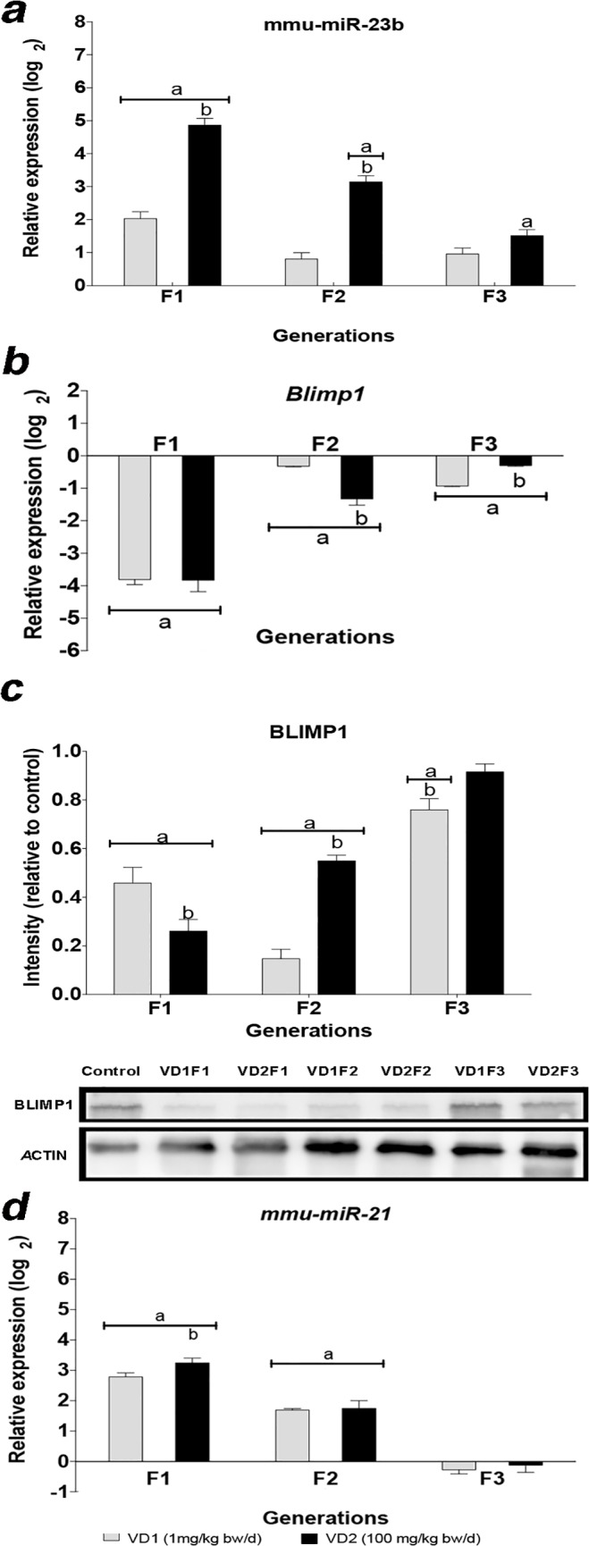
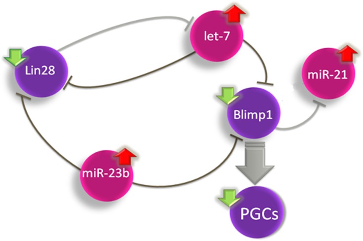
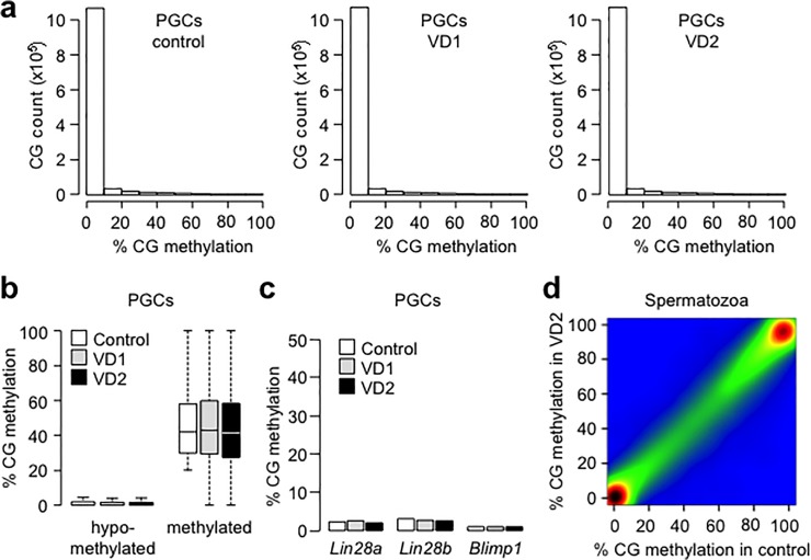
In mammals, germ cell differentiation is initiated in the Primordial Germ Cells (PGCs) during fetal development. Prenatal exposure to environmental toxicants such as endocrine disruptors may alter PGC differentiation, development of the male germline and induce transgenerational epigenetic disorders. The anti-androgenic compound vinclozolin represents a paradigmatic example of molecule causing transgenerational effects on germ cells. We performed prenatal exposure to vinclozolin in mice and analyzed the phenotypic and molecular changes in three successive generations. A reduction in the number of embryonic PGCs and increased rate of apoptotic cells along with decrease of fertility rate in adult males were observed in F1 to F3 generations. Blimp1 is a crucial regulator of PGC differentiation. We show that prenatal exposure to vinclozolin deregulates specific microRNAs in PGCs, such as miR-23b and miR-21, inducing disequilibrium in the Lin28/let-7/Blimp1 pathway in three successive generations of males. As determined by global maps of cytosine methylation, we found no evidence for prominent changes in DNA methylation in PGCs or mature sperm. Our data suggest that embryonic exposure to environmental endocrine disruptors induces transgenerational epigenetic deregulation of expression of microRNAs affecting key regulatory pathways of germ cells differentiation.


Conflict of interest statement

Competing Interests: The authors have declared that no competing interests exist.

Figures

Fig 1
Fig 1. Fertility and histopathological analysis in the testis of mice exposed to VCZ.
a) Fertility rate after fetal exposure to the low dose (VD1) or the high dose (VD2) of VCZ, expressed as a percentage of fertile males along the three generations (F1, F2 and F3). b-e) Histological analysis of testis sections stained by hematoxilyn-eosin from 10 weeks old mice from the control group (b) or VCZ exposed group (c-e), show examples of impairment of seminiferous epithelium tubule (c), tubule disintegration with cells in the lumen (d), and hypertrophic cells with fragmented karyoplasm (e, red arrows).
Fig 2
Fig 2. Apoptosis in adult testis and PGCs of mice exposed to VCZ.
a) Number of apoptotic cells counted per 10 tubules in the adult testis of control, VD1 and VD2 in F1 to F3 generations. b-d) Examples of apoptosis by TUNEL in testis sections of control (b), VD1 (c) and VD2 (d) animals. e) TUNEL positive signals after DNAse treatment of testis sections is shown as a positive control. f) Total number of PGCs isolated per testis by cell sorting with the surface marker SSEA-1 from control, VD1 and VD2 13.5dpc embryos. g) Histological evaluation of the number of PGCs in testis of 13.5dpc embryo by immunostaining with the marker SSEA-1. h) Evaluation of apoptosis in 13.5 dpc testis by TUNEL assay. i-k) Examples of co-detection of apoptosis by TUNEL and SSEA-1 positive PGCs cells by confocal microscopy analysis. i) Apoptosis detected in a PGC. j) Apoptosis detected in a somatic cell. k) None apoptotic cell detected. In the histograms, (a) indicates a significant statistical difference compared to the control value (p<0.01), and the error bars represent the standard deviation (SD).
Fig 3
Fig 3. Expression of pri-let-7a, let-7a-1-3p and Lin28 in PGCs of mice exposed to VCZ.
a-d) The graphs show the log2 of the fold change of expression in PGCs of exposed embryos relative to the unexposed control embryos. e) LIN28 protein levels measured by Western blot in 13.5 dpc PGCs. The graph bars show the quantification of protein levels normalized to the unexposed control (value = 1). In the graphs the error bars represent the standard deviation (SD), (a) indicates a significant statistical difference of VD1 and VD2 compared to the control (p≤0.01), (b) indicates a significant statistical difference of VD1 compared to VD2 (p≤0.01).
Fig 4
Fig 4. Levels of expression of Blimp1 and regulatory miRNAs in PGCs of mice exposed to VCZ.
a) Relative expression of miR-23b in PGCs of exposed embryos relative to the control embryos. b-c) Relative expression of the Blimp1 mRNA (b) and BLIMP1 protein (c) in PGCs of exposed embryos relative to the control embryos. d) Relative expression of miR-21. The error bars represent the standard deviation (SD), (a) indicates a significant statistical difference of VD1 and VD2 compared to the control (p≤0.01), (b) indicates a significant statistical difference of VD1 compared to VD2 (p≤0.01).
Fig 5
Fig 5. Schematic model for the deregulation of PGC development by the Lin28/let-7/Blimp1 pathway in the PGCs from mice exposed to VCZ.
Red arrows indicate upregulation and green arrows indicate downregulation. miRNAs are illustrated in pink. A consequence of the downregulation of BLIMP1 is the reduction in the number of PGCs in embryonic testis.
Fig 6
Fig 6. DNA Methylation analysis in PGCs and spermatozoa of F1 mice exposed to VCZ.
a) Distribution of the percentages of CpG methylation measured by RRBS in 13.5 dpc PGCs from control, VD1 and VD2 exposed embryos at the F1 generation. b) Distribution of the CG methylation measured in sequences hypomethylated in normal PGCs (defined as <20% methylation in control PGCs) or partially resistant to demethylation in PGCs (defined as >20% methylation in control PGCs). c) Evaluation of the methylation status of the promoters of Lin28a, Lin28b and Blimp1 in 13.5dpc PGCs by single locus bisulfite sequencing (for Lin28a and Lin28b) and RRBS (for Blimp1). d) Pairwise comparison of CpG methylation measured in 400bp tiles in adult spermatozoa isolated from F1VD2 males compared to control males, which reveals global conservation of DNA methylation (Pearson correlation coefficient r = 1). The density of points increase from blue to dark red.

Similar articles

Cited by

References

    1. Saitou M, Yamaji M (2012) Primordial germ cells in mice. Cold Spring Harb Perspect Biol 4. - PMC - PubMed
    1. Matzuk MM (2009) LIN28 lets BLIMP1 take the right course. Dev Cell 17: 160–161. 10.1016/j.devcel.2009.07.020 - DOI - PubMed
    1. Ginsburg M, Snow MHL, McLaren A (1990) Primordial germ cells in the mouse embryo during gastrulation. Development 110: 521–528. - PubMed
    1. Richardson BE, Lehmann R (2010) Mechanisms guiding primordial germ cell migration: strategies from different organisms. Nat Rev Mol Cell Biol 11: 37–49. 10.1038/nrm2815 - DOI - PMC - PubMed
    1. Guibert S, Forne T, Weber M (2012) Global profiling of DNA methylation erasure in mouse primordial germ cells. Genome Res 22: 633–641. 10.1101/gr.130997.111 - DOI - PMC - PubMed

Publication types

MeSH terms