. 2015 Apr 21;10(4):e0122832.
doi: 10.1371/journal.pone.0122832. eCollection 2015.

New Insights in Abdominal Pain in Paroxysmal Nocturnal Hemoglobinuria (PNH): A MRI Study

Affiliations

New Insights in Abdominal Pain in Paroxysmal Nocturnal Hemoglobinuria (PNH): A MRI Study

Francesco De Cobelli et al. PLoS One. .

Abstract

Introduction: Abdominal pain in PNH has never been investigated by in-vivo imaging studies. With MRI, we aimed to assess mesenteric vessels flow and small bowel wall perfusion to investigate the ischemic origin of abdominal pain.

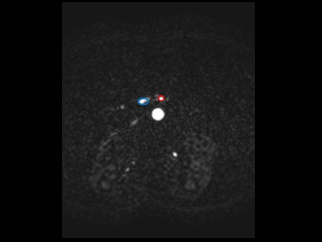
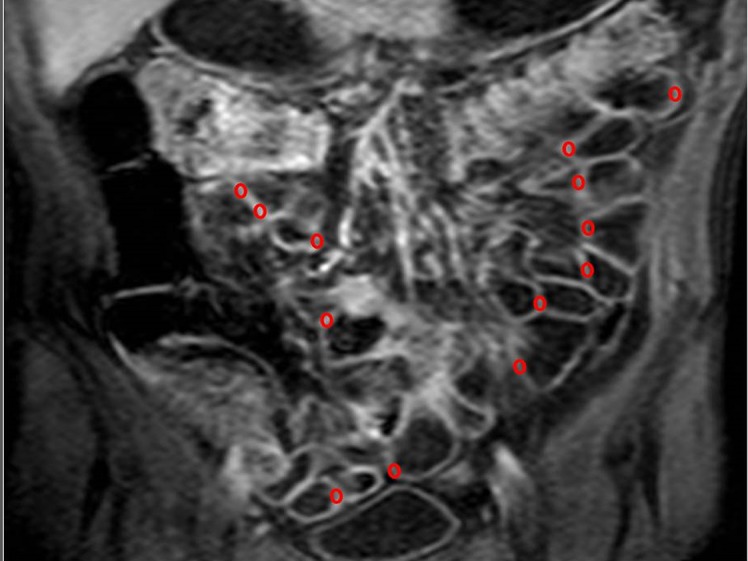
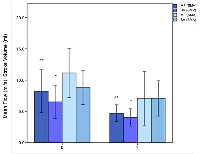
Materials and methods: Six PNH patients with (AP) and six without (NOP) abdominal pain underwent MRI. In a blinded fashion, mean flow (MF, quantity of blood moving through a vessel within a second, in mL·s-1) and stroke volume (SV, volume of blood pumped out at each heart contraction, in mL) of Superior Mesenteric Vein (SMV) and Artery (SMA), areas under the curve at 60 (AUC60) and 90 seconds (AUC90) and Ktrans were assessed by two operators.

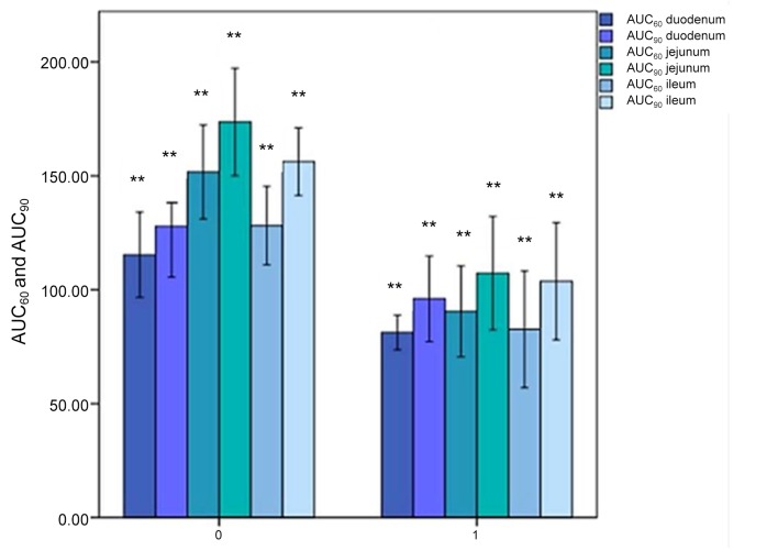
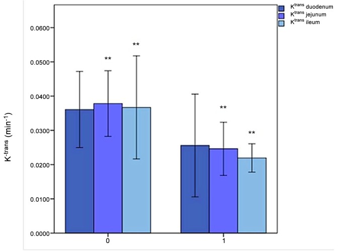
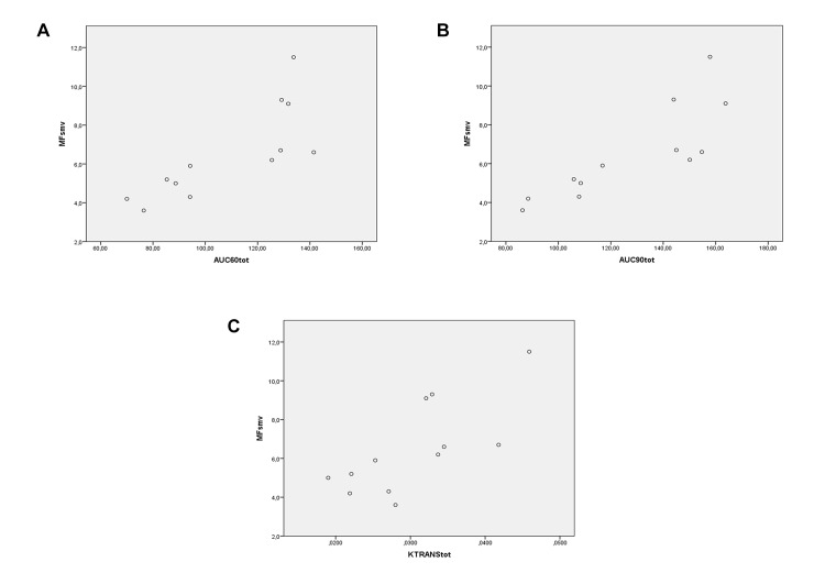
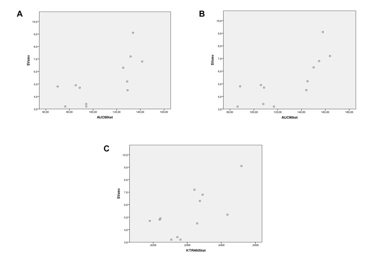
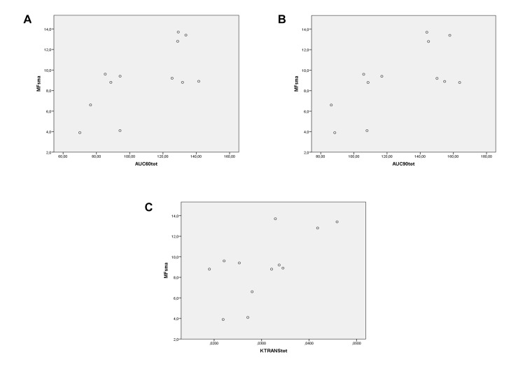
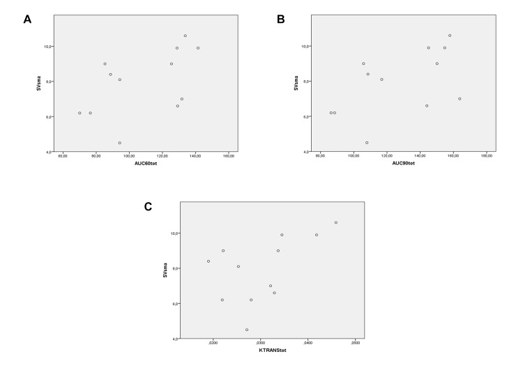
Results: Mean total perfusion and flow parameters were lower in AP than in NOP group. AUC60: 84.81 ± 11.75 vs. 131.73 ± 18.89 (P < 0.001); AUC90: 102.33 ± 14.16 vs. 152.58 ± 22.70 (P < 0.001); Ktrans: 0.0346 min-1 ± 0.0019 vs. 0.0521 ± 0.0015 (P = 0.093 duodenum, 0.009 jejunum/ileum). SMV: MF 4.67 ml/s ± 0.85 vs. 8.32 ± 2.14 (P = 0.002); SV 3.85 ml ± 0.76 vs. 6.55 ± 1.57 (P = 0.02). SMA: MF 6.95 ± 2.61 vs. 11.2 ± 2.32 (P = 0.07); SV 6.52 ± 2.19 vs. 8.78 ± 1.63 (P = 0.07). We found a significant correlation between MF and SV of SMV and AUC60 (MF:ρ = 0.88, P < 0.001; SV: ρ = 0.644, P = 0.024), AUC90 (MF: ρ = 0.874, P < 0.001; SV:ρ = 0.774, P = 0.003) and Ktrans (MF:ρ = 0.734, P = 0.007; SV:ρ = 0.581, P = 0.047).

Conclusions: Perfusion and flow MRI findings suggest that the impairment of small bowel blood supply is significantly associated with abdominal pain in PNH.


Conflict of interest statement

Competing Interests: The authors have declared that no competing interests exist.

Figures

Fig 1
Fig 1. Assessment of mean flow (MF) and stroke volume (SV).
In Phase-contrast sequences, elliptic ROIs were manually positioned on the superior mesenteric vein (SMV) and artery (SMA) in order to obtain curves of velocity and flow rate versus time.
Fig 2
Fig 2. Assessment of small bowel wall perfusion.
To calculate MRI perfusion parameters, AUC60, AUC90 and Ktrans, elliptic ROIs were positioned in different segments of the small bowel wall, particularly on proximal and distal jejunum and proximal, middle and distal ileum, as showed in this figure.
Fig 3
Fig 3. AUC60 and AUC90 in duodenum, jejunum and ileum.
The horizontal axis represents the patients without (0) and with (1) abdominal pain; the vertical axis represents the AUC60 (darker colors) and AUC90 (lighter colors) values in duodenum (blue), jejunum (green) and ileum (cyan). **: P<.01; Errors bars: 95% of confidence interval.
Fig 4
Fig 4. Ktrans in duodenum, jejunum and ileum.
The horizontal axis represents the PNH patients without (0) and with (1) abdominal pain; the vertical axis represents the Ktrans values (min-1) in duodenum, jejunum and ileum. **: P<.01; Errors bars: 95% of confidence interval.
Fig 5
Fig 5. MF and SV in the mesenteric venous (SMV) and arterial (SMA) compartment.
The horizontal axis represents the PNH patients without (0) and with (1) abdominal pain; the vertical axis represents the MF and SV values on superior mesenteric vein (SMV, dark blue) and artery (SMA, light blue). *: P<.05; **: P<.01; Errors bars: 95% of confidence interval.
Fig 6
Fig 6. Spearman rank correlation analysis.
Correlation between MRI perfusion parameters (on the horizontal axis)—AUC60 (A), AUC90 (B), and Ktrans (C)—in the whole small bowel—of PNH patients and MF of SMV (on the vertical axis).
Fig 7
Fig 7. Spearman rank correlation analysis.
Correlation between MRI perfusion parameters (on the horizontal axis)—AUC60 (A), AUC90 (B), and Ktrans (C)—in the whole small bowel—of PNH patients and SV of SMV (on the vertical axis).
Fig 8
Fig 8. Spearman rank correlation analysis.
Correlation between MRI perfusion parameters (on the horizontal axis)—AUC60 (A), AUC90 (B), and Ktrans (C)—in the whole small bowel—of PNH patients and MF of SMA (on the vertical axis).
Fig 9
Fig 9. Spearman rank correlation analysis.
Correlation between MRI perfusion parameters (on the horizontal axis)—AUC60 (A), AUC90 (B), and Ktrans (C)—in the whole small bowel—of PNH patients and SV of SMA (on the vertical axis).

Similar articles

Cited by

References

    1. Hernández-Campo PM, Almeida J, Sánchez ML, Malvezzi M, Orfao A. Normal patterns of expression of glycosylphosphatidylinositol-anchored proteins on different subsets of peripheral blood cells: a frame of reference for the diagnosis of paroxysmal nocturnal hemoglobinuria. Cytometry B Clin Cytom. 2006;70: 71–81. - PubMed
    1. Holers VM. The spectrum of complement alternative pathway-mediated diseases. Immunol Rev. 2008;223: 300–316. 10.1111/j.1600-065X.2008.00641.x - DOI - PubMed
    1. Hill A, Kelly RJ, Hillmen P. Thrombosis in paroxysmal nocturnal hemoglobinuria. Blood. 2013;121: 4985–4996. 10.1182/blood-2012-09-311381 - DOI - PubMed
    1. Wang H, Chuhjo T, Yasue S, Omine M, Nakao S. Clinical significance of a minor population of paroxysmal nocturnal hemoglobinuria-type cells in bone marrow failure syndrome. Blood. 2002;100: 3897–3902. - PubMed
    1. Gralnick HR, Vail M, McKeown LP, Merryman P, Wilson O, Chu I, et al. Activated platelets in paroxysmal nocturnal hemoglobinuria. Br J Haematol. 1995;91: 697–702. - PubMed

Publication types

MeSH terms