. 2015 Apr 21;10(4):e0120093.
doi: 10.1371/journal.pone.0120093. eCollection 2015.

Piccolo Directs Activity Dependent F-Actin Assembly from Presynaptic Active Zones via Daam1

Affiliations

Piccolo Directs Activity Dependent F-Actin Assembly from Presynaptic Active Zones via Daam1

Dhananjay Wagh et al. PLoS One. .

Abstract

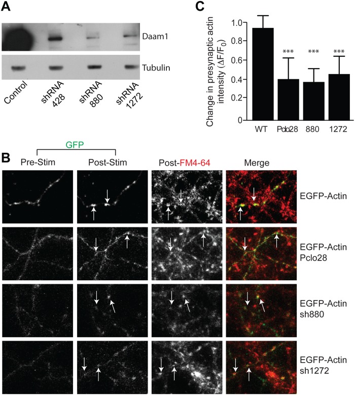
The dynamic assembly of filamentous (F) actin plays essential roles in the assembly of presynaptic boutons, the fusion, mobilization and recycling of synaptic vesicles (SVs), and presynaptic forms of plasticity. However, the molecular mechanisms that regulate the temporal and spatial assembly of presynaptic F-actin remain largely unknown. Similar to other F-actin rich membrane specializations, presynaptic boutons contain a set of molecules that respond to cellular cues and trans-synaptic signals to facilitate activity-dependent assembly of F-actin. The presynaptic active zone (AZ) protein Piccolo has recently been identified as a key regulator of neurotransmitter release during SV cycling. It does so by coordinating the activity-dependent assembly of F-Actin and the dynamics of key plasticity molecules including Synapsin1, Profilin and CaMKII. The multidomain structure of Piccolo, its exquisite association with the AZ, and its ability to interact with a number of actin-associated proteins suggest that Piccolo may function as a platform to coordinate the spatial assembly of F-actin. Here we have identified Daam1, a Formin that functions with Profilin to drive F-actin assembly, as a novel Piccolo binding partner. We also found that within cells Daam1 activation promotes Piccolo binding, an interaction that can spatially direct the polymerization of F-Actin. Moreover, similar to Piccolo and Profilin, Daam1 loss of function impairs presynaptic-F-actin assembly in neurons. These data suggest a model in which Piccolo directs the assembly of presynaptic F-Actin from the AZ by scaffolding key actin regulatory proteins including Daam1.


Conflict of interest statement

Competing Interests: The authors have declared that no competing interests exist.

Figures

Fig 1
Fig 1. Daam1 is a novel binding partner of Piccolo.
(A) Silver stained SDS PAGE gel of Piccolo antibody immunoprecipitation from P4 rat brain light membrane fraction reveals the presence of a 120 KDa band that was identified as Daam1 by mass spectrometry. (B) Western Blot analyses of similar SDS PAGE gels confirm the presence Daam1 in Piccolo immunoprecipitated fractions. Note that Bassoon, a known binding partner of Piccolo, is also immunoprecipitated while two other presynaptic proteins, Synapsin and Synaptophysin are not.
Fig 2
Fig 2. Daam1 is present within the presynaptic bouton.
(A) Western blot analysis of fractions generated by differential centrifugation of adult brain lysate demonstrates the presence of Daam1 in synaptosomes (SYN) and to a lesser extent synaptic junctions (SJ). Signals with antibodies to known synaptic junctional components (Bassoon, Piccolo, CaMKII and PSD-95) versus a synaptic vesicle protein (Synaptophysin) demonstrate the integrity of the preparation. PNS—Post nuclear supernatant, S2- post hypotonic lysis supernatant, P2- post-hypotonic lysis pellet, SYN—synaptosomes, SJ—synaptic junctions. (B) Immmunostaining of cultured hippocampal neurons (16 DIV) with antibodies against Daam1 (green) and Piccolo (red) reveals a synaptic colocalization of the two endogenous proteins. Scale bar is 5 μm. (C) Axons from EGFP-Daam1 (green) expressing neuron (14 DIV, transfected at DIV 0) immunostained with antibodies against Piccolo (red) and MAP2 (blue) reveal EGFP-Daam1 puncta within Piccolo positive presynaptic boutons juxtaposed to MAP2 positive dendrites. Scale bar is 5 μm.
Fig 3
Fig 3. Daam1 interacts with the central region of Piccolo.
(A) Schematic diagram of Piccolo depicting relative positions of different subdomains and composition of EGFP-tagged cDNA clones used in pull-down assays. (B) Western blot analysis of EGFP-tagged Piccolo fragments and Myc-Daam1 expressed and immunoprecipitated from COS7 cells with an antibody to GFP. (C) Schematic diagram of Daam1 depicting relative positions of different subdomains and organization of Myc-tagged cDNA clones. (D) Western blot analysis of Myc-tagged Daam1 constructs co-expressed with EGFP-Pclo1980-2553 and immunoprecipitated from COS7 cells with an antibody to Piccolo. Q—polyQ domain; Zn1 and Zn2—zinc fingers 1 and 2; CC—coiled coil domain; PRS—proline rich sequence; PDZ—PDZ domain; C2—C2 domain; GBD—Rho GTPase binding domain; DID—Diaphanous Inhibitory Domain; FH1 and FH2—formin homology domains 1 and 2; DAD—Diaphanous Autoregulatory Domain.
Fig 4
Fig 4. Pclo1980-2553 localizes to stress fibers induced by activated Daam1.
Expression and immunostaining of COS7 cells transfected Myc tagged Daam1 isoforms (red) alone (A) or co-expresssed (B) with EGFP or EGFP-Pclo1980-2553 (green). Alexa Fluor-conjugated phalloidin (blue) identifies actin rich structures including stress fibers. The scale bars are both 5 μm and apply to all images in the respective panels.
Fig 5
Fig 5. Plasma membrane targeting of Pclo1980-2553 accentuates association with stress fibers induced by activated Daam1.
(A) Single plane confocal images of COS7 cells expressing CD4-EGFP-Pclo1980-2553 chimera (green) and Myc-C-Daam1 (not displayed), treated with anti-CD4 antibodies to patch surface CD4 fusion proteins, and then, labeled with Alexa-Fluor 568-conjugated phalloidin (red). (B) As in (A), but with CD4-EGFP instead of CD4-EGFP-Pclo1980-2553 chimera. Note the presence of stress-fibers in A and B is indicative of Myc-C-Daam1 expression (see Fig 4).
Fig 6
Fig 6. Pclo1980-2553 serves as a platform for Daam1 dependent F-actin assembly.
(A) Discrete round structures demonstrate that CD4-EGFP-Pclo1980-2553 accumulates around CD4 antibody-coupled Protein-A beads dropped onto COS7 cells. Scale bar is 20 μm. (B) When coexpressed with CD4-EGFP-Pclo1980-2553 (top panels, green in merge) Myc-C-Daam1 (right middle panel, red in merge), but not full length Myc-Daam1 (left middle panel, red in merge), is recruited to CD4 antibody-coupled Protein A bead foci. (C) Visualization of F-actin accumulation by AlexaFluor 568-conjugated Phalloidin (Red) around CD4 antibody-coupled beads dropped onto COS7 cells expressing CD4-EGFP or CD4-EGFP-Pclo1980-2553 alone (top panels), CD4-EGFP or CD4-EGFP-Pclo1980-2553 with Myc-Daam1 (Middle panels) or CD4-EGFP or CD4-EGF-Pclo1980-2553 with Myc-C-Daam1 (bottom panels). (D) Quantitation of relative accumulation of phalloidin intensity around CD4 antibody-coupled beads in COS7 cells expressing the indicated constructs. Data are expressed as mean with error bars representing standard deviation. For statistical analysis, within each group (no Daam1, Daam1, and C-Daam1) a comparison was made between cells transfected with CD4-EGFP and those transfected with CD4-EGFP-Pclo1980-2553 using a two-tailed t-test (** p<0.01). (E) Co-expression of mRFP-UtrophinCHD allows visualization of active F-actin assembly (bottom panels, red in merge) around CD4 antibody-coupled beads in COS7 cells expressing Myc-C-Daam1 and CD4-EGFP-Pclo1980-2553 (top left), but not CD4-EGFP (top right). (F) Time lapse image sequence of COS7 cells expressing Myc-C-Daam1 (not displayed), mRFP-UtrophinCHD (Red) and CD4-EGFP (top panels) or CD4-EGFP-Pclo1980-2553 (bottom panels) treated with 5 μM Latrunculin-A 5 min prior to the addition of CD4-antibody coated beads. Continued accumulation of mRFP-UtrophinCHD at beads on cells expressing Myc-C-Daam1 and CD4-EGFP-Pclo1980-2553 in the presence of 5 μM Latrunculin-A is indicative of the potent induction of F-actin assembly by these molecules. Scales bars in (B), (C), (E), and (F) are 5 μm.
Fig 7
Fig 7. Pclo1980-2553 is targeted into filopodia when co-expressed with activated Daam1.
(A) Labeling of mitochondria with Mitotracker Green (upper panels, green in merge) in COS7 cells expressing ActA-mRFP or ActA-Pclo1980-2553-mRFP (middle panels, red in merge) demonstrates localization of Pclo1980-2553 sequences to the surface of mitochondria. (B) Expression of ActA-mRFP (top set of panels, red in merge) or ActA-Pclo1980-2553-mRFP (bottom set of panels, red in merge) with Myc-C-Daam1 (green in merge) along with Actin labeling with Alexa fluor-coupled Phalloidin (blue in merge) reveals the accumulation of ActA-Pclo1980-2553-mRFP labeled mitochondria in phalloidin positive filopodia. (C) Analysis of mitochondrial expression pattern indicates nearly all cells expressing ActA-Pclo1980-2553-mRFP demonstrate accumulation in filopodia while this pattern is seen in less than half of the cells expressing ActA-mRFP. Data are expressed as mean with error bars representing standard deviation. For statistical analysis, comparison was made between the control cells (those expressing Mcy-C-Daam1 with ActA) and cells expressing Mcy-C-Daam1 with ActA coupled to Pclo1980-2553 using a two-tailed t-test (* p<0.05).
Fig 8
Fig 8. Daam1 is required for activity dependent assembly of presynaptic F-actin.
(A) Western blot analysis of extracts (5 μg total protein) from cultured hippocampal neurons (DIV15) infected with lentiviral vectors expressing EGFP-Actin alone (control) or with the indicated shRNA targeting Daam1 (sh428, sh880, or sh1272). Antibodies against Daam1 confirm that the shRNAs markedly reduce expression of the protein relative to a control protein (tubulin). Analysis of neomycin expression from the plasmid confirms similar levels of transfection efficiencies. (B) Live cell fluorescent images of cultured hippocampal neurons (14DIV), infected with lentiviral vectors expressing EGFP-Actin (green) alone or with shRNA against Piccolo (Pclo28) or Daam1 (sh880 or sh1272). Cells were stimulated with 90mM KCl for 60 sec in the presence of FM4-64 (red) to promote the assembly of presynaptic F-actin and detect presynaptic sites capable of recycling their synaptic vesicles pools. EGFP-Actin expressed alone (top panels) readily accumulates at FM4-64 dye uptake sites, while only modest accumulation is observed in neurons lacking Piccolo (Pclo28 labeled panels) or Daam1 (sh880, sh1272 labeled panels). Arrows indicate examples of co-localization of EGFP-Actin and FM4-64 positive puncta post stimulation. (C) Quantitation of the change in EGFP-Actin fluorescence at the presynaptic boutons measured pre-and post stimulation at the FM dye uptake sites in presence of Piccolo (TS28) or Daam1 (sh1272, sh880) shRNAs demonstrates a marked decrease in Actin clustering with stimulation when either protein is targeted. Data are expressed as mean with error bars representing standard deviation. For statistical analysis, comparison was made between the control and the specific shRNA samples using a two-tailed t-test (*** p<0.001).

Similar articles

Cited by

References

    1. Sudhof TC. The synaptic vesicle cycle. Annu Rev Neurosci. 2004;27:509–47. . - PubMed
    1. Schoch S, Gundelfinger ED. Molecular organization of the presynaptic active zone. Cell Tissue Res. 2006;326(2):379–91. . - PubMed
    1. Rosenmund C, Rettig J, Brose N. Molecular mechanisms of active zone function. Curr Opin Neurobiol. 2003;13(5):509–19. . - PubMed
    1. Greengard P, Benfenati F, Valtorta F. Synapsin I, an actin-binding protein regulating synaptic vesicle traffic in the nerve terminal. Adv Second Messenger Phosphoprotein Res. 1994;29:31–45. Epub 1994/01/01. . - PubMed
    1. Hilfiker S, Pieribone VA, Czernik AJ, Kao HT, Augustine GJ, Greengard P. Synapsins as regulators of neurotransmitter release. Philos Trans R Soc Lond B Biol Sci. 1999;354(1381):269–79. . - PMC - PubMed

Publication types

MeSH terms

LinkOut - more resources