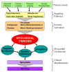
Myocardial factor revisited: The importance of myocardial fibrosis in adults with congenital heart disease
- PMID: 25897907
- PMCID: PMC4446173
- DOI: 10.1016/j.ijcard.2015.04.064
Myocardial factor revisited: The importance of myocardial fibrosis in adults with congenital heart disease
Abstract
Pioneers in congenital heart surgery observed that exercise capacity did not return to normal levels despite successful surgical repair, leading some to cite a "myocardial factor" playing a role. They conjectured that residual alterations in myocardial function would be significant for patients' long-term outlook. In fulfillment of their early observations, today's adult congenital heart disease (ACHD) population shows well-recognized features of heart failure, even among patients without clear residual anatomic or hemodynamic abnormalities, demonstrating the vital role of the myocardium in their morbidity and mortality. Whereas the 'myocardial factor' was an elusive concept in the early history of congenital heart care, we now have imaging techniques to detect and quantify one such factor--myocardial fibrosis. Understanding the importance of myocardial fibrosis as a final common pathway in a variety of congenital lesions provides a framework for both the study and treatment of clinical heart failure in this context. While typical heart failure pharmacology should reduce or attenuate fibrogenesis, efforts to show meaningful improvements with standard pharmacotherapy in ACHD repeatedly fall short. This paper considers the importance of myocardial fibrosis and function, the current body of evidence for myocardial fibrosis in ACHD, and its implications for research and treatment.
Keywords: Cardiac magnetic resonance; Congenital; Heart defects; Heart failure; Myocardial fibrosis.
Copyright © 2015 Elsevier Ireland Ltd. All rights reserved.
Conflict of interest statement
Neither author has any conflicts of interest related to this topic.
Figures


References
-
- Warnes CA, Liberthson R, Danielson GK, Dore A, Harris L, Hoffman JI, Somerville J, Williams RG, Webb GD. Task force 1: The changing profile of congenital heart disease in adult life. J Am Coll Cardiol. 2001;37:1170–1175. - PubMed
-
- Marelli AJ, Mackie AS, Ionescu-Ittu R, Rahme E, Pilote L. Congenital heart disease in the general population: Changing prevalence and age distribution. Circulation. 2007;115:163–172. - PubMed
-
- Blalock A. Cardiovascular surgery, past and present. J Thorac Cardiovasc Surg. 1966;51:153–167. - PubMed
-
- McIntosh HD, Cohen AI. Pulmonary stenosis; the importance of the myocardial factor in determining the clinical course and surgical results. Am Heart J. 1963;65:715–716.
-
- Goldberg SJ, Weiss R, Adams FH. A comparison of the maximal endurance of normal children and patients with congenital cardiac disease. The Journal of pediatrics. 1966;69:46–55. - PubMed
Publication types
MeSH terms
Grants and funding
LinkOut - more resources
Full Text Sources
Other Literature Sources
Medical

