

EGFR inhibitors prevent induction of cancer stem-like cells in esophageal squamous cell carcinoma by suppressing epithelial-mesenchymal transition
- PMID: 25897987
- PMCID: PMC4623069
- DOI: 10.1080/15384047.2015.1040959
EGFR inhibitors prevent induction of cancer stem-like cells in esophageal squamous cell carcinoma by suppressing epithelial-mesenchymal transition
Abstract
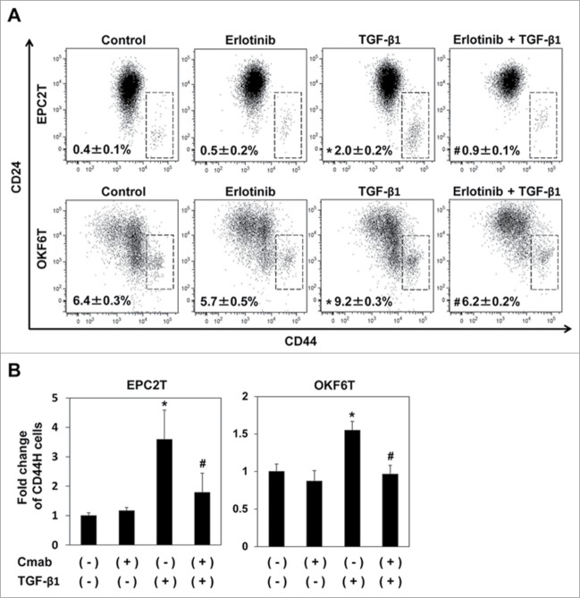
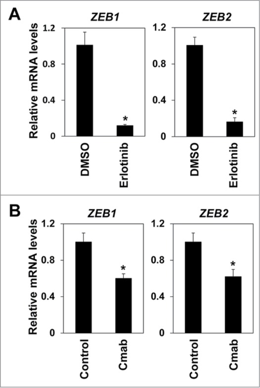
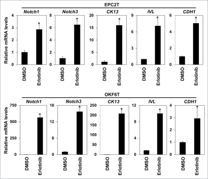
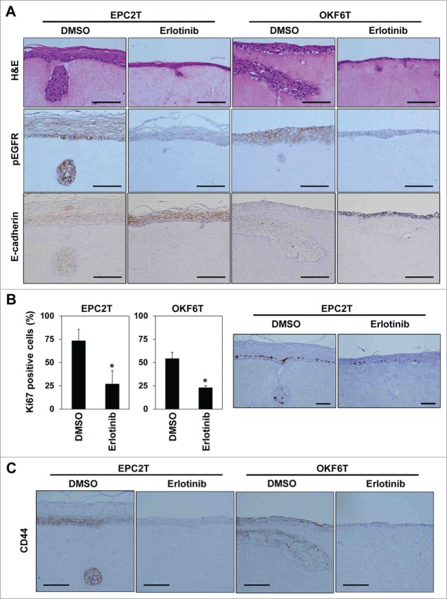
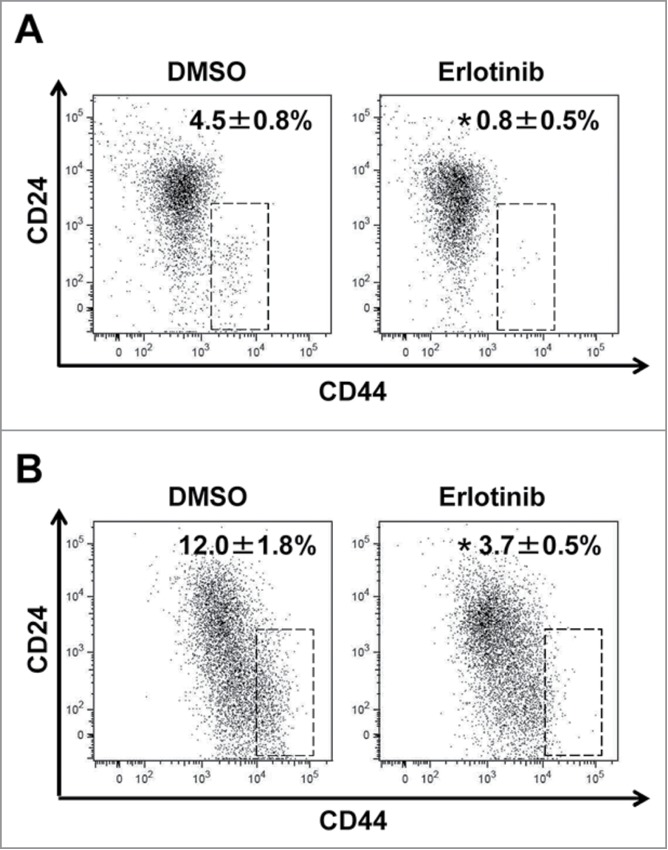
There exists a highly tumorigenic subset of esophageal squamous cell carcinoma (ESCC) cells defined by high expression of CD44. A novel therapy targeting these cancer stem-like cells (CSCs) is needed to improve prognosis of ESCC. CSCs of ESCC have a mesenchymal phenotype and epithelial-mesenchymal transition (EMT) is critical to enrich and maintain CSCs. EGFR, frequently overexpressed in ESCC, has pivotal roles in EMT induced by TGF-β in invasive fronts. Thus, EMT in invasive fronts of ESCC might be important for CSCs and EGFR could be a target of a novel therapy eliminating CSCs. However, effects of EGFR inhibitors on CSCs in ESCC have not been fully examined. EGFR inhibitors, erlotinib and cetuximab, significantly suppressed enrichment of CSCs via TGF-β1-mediated EMT. Importantly, EGFR inhibitors sharply suppressed ZEB1 that is essential for EMT in ESCC. Further, EGFR inhibitors activated Notch1 and Notch3, leading to squamous cell differentiation. EGFR inhibition may suppress expression of ZEB1 and induce differentiation, thereby blocking EMT-mediated enrichment of CSCs. In organotypic 3D culture, a form of human tissue engineering, tumor cells in invasive nests showed high expression of CD44. Erlotinib significantly blocked invasion into the matrix and CD44 high expressing CSCs were markedly suppressed by erlotinib in organotypic 3D culture. In conclusion, EMT is a critical process for generation of CSCs and the invasive front of ESCC, where EMT occurs, might form a CSC niche in ESCC. EGFR inhibitors could suppress EMT in invasive fronts and be one therapeutic option targeting against generation of CSCs in ESCC.
Keywords: CK13, cytokeratin 13; CSCs, cancer stem-like cells; DMSO, dimethyl sulfoxide; EMT, epithelial–mesenchymal transition; EGFR, epidermal growth factor receptor; ESCC, esophageal squamous cell carcinoma; FACS, fluorescence-activated cell sorting; IHC, immun; EGFR inhibitor; ZEB1; cancer stem cell; epithelial-mesenchymal transition; esophageal squamous cell carcinoma; organotypic 3D culture.
Figures
Similar articles
-
Distinct effects of EGFR inhibitors on epithelial- and mesenchymal-like esophageal squamous cell carcinoma cells.J Exp Clin Cancer Res. 2017 Aug 1;36(1):101. doi: 10.1186/s13046-017-0572-7. J Exp Clin Cancer Res. 2017. PMID: 28764725 Free PMC article.
-
Fibroblast growth factor-2-mediated FGFR/Erk signaling supports maintenance of cancer stem-like cells in esophageal squamous cell carcinoma.Carcinogenesis. 2017 Oct 26;38(11):1073-1083. doi: 10.1093/carcin/bgx095. Carcinogenesis. 2017. PMID: 28927233 Free PMC article.
-
Programmed death-ligand 1 expression at tumor invasive front is associated with epithelial-mesenchymal transition and poor prognosis in esophageal squamous cell carcinoma.Cancer Sci. 2017 Jun;108(6):1119-1127. doi: 10.1111/cas.13237. Epub 2017 May 25. Cancer Sci. 2017. PMID: 28294486 Free PMC article.
-
Linking Cancer Stem Cell Plasticity to Therapeutic Resistance-Mechanism and Novel Therapeutic Strategies in Esophageal Cancer.Cells. 2020 Jun 17;9(6):1481. doi: 10.3390/cells9061481. Cells. 2020. PMID: 32560537 Free PMC article. Review.
-
Cancer stem cells in esophageal squamous cell carcinoma.Pathol Res Pract. 2022 Sep;237:154043. doi: 10.1016/j.prp.2022.154043. Epub 2022 Jul 30. Pathol Res Pract. 2022. PMID: 35926434 Review.
Cited by
-
Tumor-associated macrophage-derived cytokines enhance cancer stem-like characteristics through epithelial-mesenchymal transition.Onco Targets Ther. 2018 Jul 4;11:3817-3826. doi: 10.2147/OTT.S168317. eCollection 2018. Onco Targets Ther. 2018. PMID: 30013362 Free PMC article. Review.
-
Nimotuzumab Plus Paclitaxel and Cisplatin as a 1st-Line Treatment for Esophageal Cancer: Long Term Follow-up of a Phase II Study.J Cancer. 2019 Feb 23;10(6):1409-1416. doi: 10.7150/jca.28659. eCollection 2019. J Cancer. 2019. PMID: 31031851 Free PMC article.
-
Clinical and Prognostic Implications of P21 (WAF1/CIP1) Expression in Patients with Esophageal Cancer: A Systematic Review and Meta-Analysis.Dis Markers. 2020 Jan 7;2020:6520259. doi: 10.1155/2020/6520259. eCollection 2020. Dis Markers. 2020. PMID: 31998417 Free PMC article.
-
Whole-Genome Sequencing Reveals Diverse Models of Structural Variations in Esophageal Squamous Cell Carcinoma.Am J Hum Genet. 2016 Feb 4;98(2):256-74. doi: 10.1016/j.ajhg.2015.12.013. Epub 2016 Jan 28. Am J Hum Genet. 2016. PMID: 26833333 Free PMC article.
-
MicroRNAs as Epigenetic Determinants of Treatment Response and Potential Therapeutic Targets in Prostate Cancer.Cancers (Basel). 2021 May 14;13(10):2380. doi: 10.3390/cancers13102380. Cancers (Basel). 2021. PMID: 34069147 Free PMC article. Review.
References
-
- Pennathur A, Gibson MK, Jobe BA, Luketich JD. Oesophageal carcinoma. Lancet 2013; 381:400-12; PMID:23374478; http://dx.doi.org/10.1016/S0140-6736(12)60643-6 - DOI - PubMed
-
- Fokas E, Weiss C, Rodel C. The role of radiotherapy in the multimodal management of esophageal cancer. Dig Dis 2013; 31:30-7; PMID:23797120; http://dx.doi.org/10.1159/000347170 - DOI - PubMed
-
- Oyama T, Tomori A, Hotta K, Morita S, Kominato K, Tanaka M, Miyata Y. Endoscopic submucosal dissection of early esophageal cancer. Clin Gastroenterol Hepatol 2005; 3:S67-70; PMID:16013002; http://dx.doi.org/10.1016/S1542-3565(05)00291-0 - DOI - PubMed
-
- Shimizu Y, Tsukagoshi H, Fujita M, Hosokawa M, Kato M, Asaka M. Long-term outcome after endoscopic mucosal resection in patients with esophageal squamous cell carcinoma invading the muscularis mucosae or deeper. Gastrointest Endosc 2002; 56:387-90; PMID:12196777; http://dx.doi.org/10.1016/S0016-5107(02)70043-6 - DOI - PubMed
-
- Shaheen NJ, Sharma P, Overholt BF, Wolfsen HC, Sampliner RE, Wang KK, Galanko JA, Bronner MP, Goldblum JR, Bennett AE, et al.. Radiofrequency ablation in Barrett's esophagus with dysplasia. N Engl J Med 2009; 360:2277-88; PMID:19474425; http://dx.doi.org/10.1056/NEJMoa0808145 - DOI - PubMed
Publication types
MeSH terms
Substances
Grants and funding
LinkOut - more resources
Full Text Sources
Other Literature Sources
Medical
Research Materials
Miscellaneous