Review
. 2015 Apr 17;6(2):216-37.
doi: 10.3390/genes6020216.

Genetic mosaics and the germ line lineage

Affiliations
Review

Genetic mosaics and the germ line lineage

Mark E Samuels et al. Genes (Basel). .

Abstract

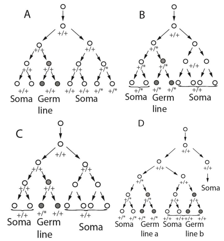
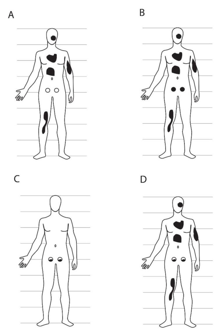
Genetic mosaics provide information about cellular lineages that is otherwise difficult to obtain, especially in humans. De novo mutations act as cell markers, allowing the tracing of developmental trajectories of all descendants of the cell in which the new mutation arises. De novo mutations may arise at any time during development but are relatively rare. They have usually been observed through medical ascertainment, when the mutation causes unusual clinical signs or symptoms. Mutational events can include aneuploidies, large chromosomal rearrangements, copy number variants, or point mutations. In this review we focus primarily on the analysis of point mutations and their utility in addressing questions of germ line versus somatic lineages. Genetic mosaics demonstrate that the germ line and soma diverge early in development, since there are many examples of combined somatic and germ line mosaicism for de novo mutations. The occurrence of simultaneous mosaicism in both the germ line and soma also shows that the germ line is not strictly clonal but arises from at least two, and possibly multiple, cells in the embryo with different ancestries. Whole genome or exome DNA sequencing technologies promise to expand the range of studies of genetic mosaics, as de novo mutations can now be identified through sequencing alone in the absence of a medical ascertainment. These technologies have been used to study mutation patterns in nuclear families and in monozygotic twins, and in animal model developmental studies, but not yet for extensive cell lineage studies in humans.


Figures

Figure 1
Figure 1
Potential cell lineages of somatic and germ line mosaicism based on alternative specification scenarios. In each panel, the one cell zygote is shown at the top, and following a small number of divisions. At some point, a wild type (+/+) cell gives rise to a mutant cell (+/*), which divides to produce a set of descendants that include all or part of the germ line and/or part of the soma. Panels (A), (B), and (C) assume that a single post-zygotic cell (FGC1) gives rise to the entire germ line. Panel (D) assumes two or more post-zygotic cells, of different post-fertilization ancestry, contribute to different portions of the germ line (Germ line a, Germ line b). In each panel, cells contributing to the germ line are shaded in grey. De novo mutations are indicated as asterisks so that cells carrying the mutation are heterozygous +/*. Panel (D) shows the lineage outcome in the event of mutation in a cell that gives rise to both germ-line and somatic descendants, in the case of multiple (i.e., non-clonal) germ line precursors. These trees are not meant to reflect specific actual cell lineages or the relative size of germ line versus somatic cell compartments.
Figure 2
Figure 2
Potential anatomical patterns of mosaicism based on the lineage and mutational scenarios of Figure 1. Each panel is matched to the equivalent panel in Figure 1 (panel A, germ line clonal, mutation in soma; B, germ line clonal, mutation in cell ancestral to germ line and some of soma; C, germ line clonal, mutation in germ line; D, germ line not clonal, mutation in cell ancestral to some of germ line and some of soma). The germ line is presented as two circles in the lower abdomen, either wild type (unshaded) or mutant (black). Patches of somatic tissue carrying a de novo mutation are shown as irregular black shapes and may include skin and/or any other kind of somatic cells. In panels (C) and (D), germ line mosaicism is shown in both the right and left gonad, but other gonad patterns are likely as well, depending on how the primordial gonads are populated by PGCs. Relative sizes of the germ line versus soma are not to scale; the germ line in humans is obviously much smaller than the soma in physical size and cell number. Patterns of mosaicism are also not meant to be interpreted literally, other than inclusion or exclusion of all or part of germ line versus soma.

References

    1. De Felici M. Origin, Migration, and Proliferation of Human Primordial Germ Cells. In: Coticchio C., Albertini D.F., de Santis L., editors. Oogenesis. Springer-Verlag; London, UK: 2013. pp. 19–37.
    1. McKay D.G., Hertig A.T., Adams E.C., Danziger S. Histochemical observations on the germ cells of human embryos. Anat. Rec. 1953;117:201–219. doi: 10.1002/ar.1091170206. - DOI - PubMed
    1. Chiquoine A.D. The identification, origin, and migration of the primordial germ cells in the mouse embryo. Anat. Rec. 1954;118:135–146. doi: 10.1002/ar.1091180202. - DOI - PubMed
    1. Eddy E.M., Clark J.M., Gong D., Fenderson B.A. Origin and migration of primordial germ cells in mammals. Gamete Res. 1981;4:333–362. doi: 10.1002/mrd.1120040407. - DOI
    1. Hardisty M.W. The numbers of vertebrate primordial germ cells. Biol. Rev. Camb. Philos. Soc. 1967;42:265–287. doi: 10.1111/j.1469-185X.1967.tb01420.x. - DOI - PubMed