. 2015 Aug 21;36(32):2184-96.
doi: 10.1093/eurheartj/ehv109. Epub 2015 Apr 21.

Osteopontin is indispensible for AP1-mediated angiotensin II-related miR-21 transcription during cardiac fibrosis

Affiliations

Osteopontin is indispensible for AP1-mediated angiotensin II-related miR-21 transcription during cardiac fibrosis

Johan M Lorenzen et al. Eur Heart J. .

Abstract

Aims: Osteopontin (OPN) is a multifunctional cytokine critically involved in cardiac fibrosis. However, the underlying mechanisms are unresolved. Non-coding RNAs are powerful regulators of gene expression and thus might mediate this process.

Methods and results: OPN and miR-21 were significantly increased in cardiac biopsies of patients with myocardial fibrosis. Ang II infusion via osmotic minipumps led to specific miRNA regulations with miR-21 being strongly induced in wild-type (WT) but not OPN knockout (KO) mice. This was associated with enhanced cardiac collagen content, myofibroblast activation, ERK-MAP kinase as well as AKT signalling pathway activation and a reduced expression of Phosphatase and Tensin Homologue (PTEN) as well as SMAD7 in WT but not OPN KO mice. In contrast, cardiotropic AAV9-mediated overexpression of OPN in vivo further enhanced cardiac fibrosis. In vitro, Ang II induced expression of miR-21 in WT cardiac fibroblasts, while miR-21 levels were unchanged in OPN KO fibroblasts. As pri-miR-21 was also increased by Ang II, we studied potential involved upstream regulators; Electrophoretic Mobility Shift and Chromatin Immunoprecipitation analyses confirmed activation of the miR-21 upstream-transcription factor AP-1 by Ang II. Recombinant OPN directly activated miR-21, enhanced fibrosis, and activated the phosphoinositide 3-kinase pathway. Locked nucleic acid-mediated miR-21 silencing ameliorated cardiac fibrosis development in vivo.

Conclusion: In cardiac fibrosis related to Ang II, miR-21 is transcriptionally activated and targets PTEN/SMAD7 resulting in increased fibroblast survival. OPN KO animals are protected from miR-21 increase and fibrosis development due to impaired AP-1 activation and fibroblast activation.

Keywords: Angiotensin II; Cardiac fibrosis; Osteopontin; miR-21; microRNA.


Figures

Figure 1
Figure 1
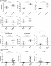
Ang II-induced inflammation and fibrosis: angiotensin II induces the mRNA expression of osteopontin (A), interleukin-6 (B), as well as macrophage chemoattractant protein-1 (C) in wild-type fibroblasts, while interleukin-6 (D) and macrophage chemoattractant protein-1 (E) levels remain unchanged in osteopontin knockout fibroblasts in response to angiotensin II. Angiotensin II induces the mRNA expression of osteopontin in vitro in cardiomyocytes (F) as well as the secretion of osteopontin in the cell-culture supernatant of cultured cardiomyocytes (G). n = 5 independent experiments. Expression of collagen I (H), osteopontin (I), as well as miR-21 (J) in cardiac biopsies of patients with myocardial fibrosis related to aortic stenosis (n = 15) compared with healthy control patients (n = 5). Concomitant angiotensin-receptor blocker treatment lowers elevated levels of osteopontin in biopsies of patients with myocardial fibrosis (K).
Figure 2
Figure 2
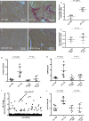
Ang II-induced fibrosis and miRNA expression in osteopontin wild type and knockout mice: Sirius red staining in paraffin-embedded sections of cardiac tissue of osteopontin wild type mice following vehicle-infusion (2 weeks, PBS, A) and Ang II infusion (B) as well as quantification of results (C) and osteopontin knockout mice following vehicle-infusion (D) and Ang II infusion (E) as well as quantification of results (F). Expression of Collagen I (G) and alpha smooth muscle actin (α-SMA, H) mRNA following Ang II or vehicle infusion. A global miRNA screen in hearts of osteopontin wild type and osteopontin knockout mice following Ang II infusion (I). MiR-21 expression in osteopontin wild type and knockout animals (J). n = 6 animals per group and analysis.
Figure 3
Figure 3
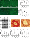
Effects of osteopontin in vivo: wheat-germ agglutintin staining in osteopontin wild type (A and B) and knockout (C and D) mice to visualize cell membranes of cardiomyocytes. Cardiomyocyte cell size was counted and quantified (E). Heart weight/body weight ratio in osteopontin wild type and knockout mice (F). Downstream signalling pathways in vivo: phosphorylation of AKT (G and H), SMAD2,3 (G and I) and ERK (G and J) in vivo. In areas of increased perivascular fibrosis (Sirus red staining, K), osteopontin expression (brown staining) is also increased (L). n = 6 animals per group and analysis.
Figure 4
Figure 4
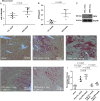
MiR-21 expression in mice following cardiotropic osteopontin-AAV9 injection (A) as well as osteopontin protein expression in hearts (B and C) compared with CTL-AAV9. Sirius red staining in paraffin-embedded sections of cardiac tissue of sham-operated mice (D), mice subjected to CTL-AAV9 and Ang II (E), osteopontin-AAV9 and Ang II (F), CTL-AAV9 and Ang II as well as locked-nucleic acid treatment targeting miR-21 (LNA-21, G), osteopontin-AAV9 and Ang II as well as LNA-21 (H), and quantification of results (I). n = 5 animals per group and analysis.
Figure 5
Figure 5
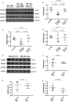
miR-21 targets in osteopontin wild type and knockout mice: Protein expression as well as densitometric quantification of PTEN (A and B), SMAD7 (A and C), and PDCD4 (A and D) in wild type mice subjected to Ang II and locked-nucleic acid treatment targeting miR-21 (LNA-21) or control mismatch LNA (LNA–MM) treatment. Protein expression as well as densitometric quantification of PTEN (E and F), SMAD7 (E and G), and PDCD4 (E and H) in osteopontin knockout mice subjected to Ang II infusion. n = 6 animals per group and analysis.
Figure 6
Figure 6
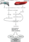
Proposed scheme of angiotensin II-induced osteopontin expression: angiotensin II leads to the expression of osteopontin in cardiac fibroblasts, which induces AP-1-mediated transcription of miR-21. Mature miR-21 targets and down-regulates PTEN, which subsequently leads to phosphorylation of AKT and nuclear exclusion of FOXO3a. In addition, miR-21 targets SMAD7, which results in phosphorylation of ERK. In cardiomyocytes, angiotensin II leads to the secretion of osteopontin, which induces the aforementioned mechanisms. Moreover, secreted osteopontin activates the PI3-kinase pathway as well as AKT phosphorylation. Taken together these mechanisms results in fibroblast survival and deposition of extracellular matrix (ECM).

Comment in

References

    1. Thum T, Lorenzen JM. Cardiac fibrosis revisited by microRNA therapeutics. Circulation 2012;126:800–802. - PubMed
    1. Wynn TA. Common and unique mechanisms regulate fibrosis in various fibroproliferative diseases. J Clin Invest 2007;117:524–529. - PMC - PubMed
    1. Mazzali M, Kipari T, Ophascharoensuk V, Wesson JA, Johnson R, Hughes J. Osteopontin – a molecule for all seasons. QJM 2002;95:3–13. - PubMed
    1. Lenga Y, Koh A, Perera AS, McCulloch CA, Sodek J, Zohar R. Osteopontin expression is required for myofibroblast differentiation. Circ Res 2008;102:319–327. - PubMed
    1. Singh K, Sirokman G, Communal C, Robinson KG, Conrad CH, Brooks WW, Bing OH, Colucci WS. Myocardial osteopontin expression coincides with the development of heart failure. Hypertension 1999;33:663–670. - PubMed

Publication types

MeSH terms