. 2015 Oct;35(7):1003-15.
doi: 10.1007/s10571-015-0195-5. Epub 2015 Apr 22.

Resveratrol Rescues the Impairments of Hippocampal Neurons Stimulated by Microglial Over-Activation In Vitro

Affiliations

Resveratrol Rescues the Impairments of Hippocampal Neurons Stimulated by Microglial Over-Activation In Vitro

Feng Wang et al. Cell Mol Neurobiol. 2015 Oct.

Abstract

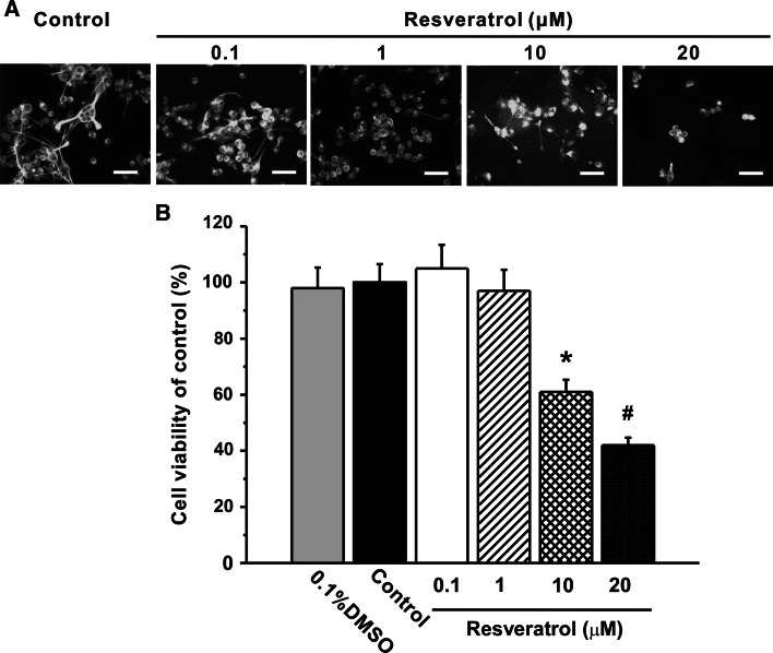
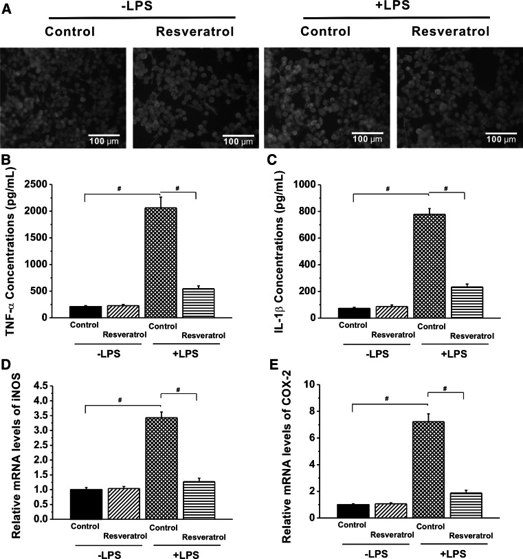
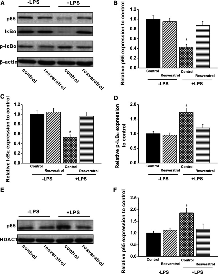
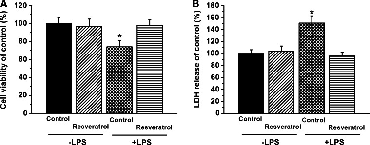
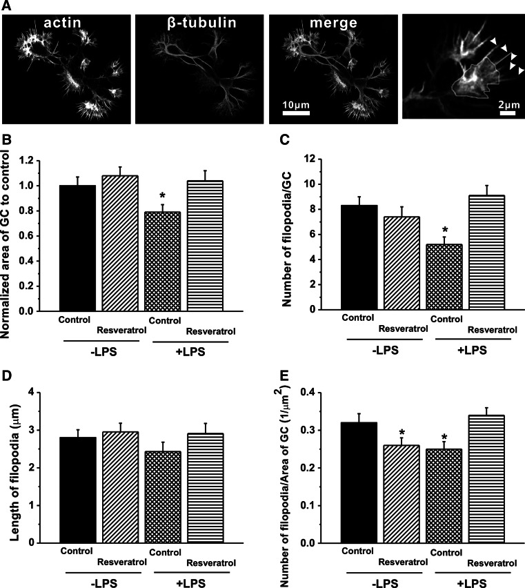
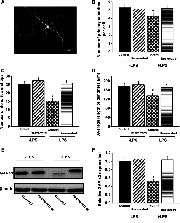
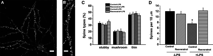
Resveratrol is a naturally occurring phytoalexin found in red grapes, and believed to have neuroprotective, anti-oxidant, and anti-inflammatory effects. But little is known about its effect on the neural impairments induced by microglial over-activation, which leads to neuroinflammation and multiple pathophysiological damages. In this study, we aimed to investigate the protective effects of resveratrol on the impairments of neural development by microglial over-activation insult. The results indicated that resveratrol inhibited the lipopolysaccharide (LPS)-dependent release of cytokines from activated microglia and LPS-dependent changes in NF-κB signaling pathway. Conditioned medium (CM) from activated microglia treated by resveratrol directly protected primary cultured hippocampal neurons against LPS-CM-induced neuronal death, and restored the inhibitory effects of LPS-CM on dendrite sprouting and outgrowth. Finally, neurons cultured in CM from LPS-stimulated microglia treated by resveratrol exhibited increased spine density compared to those without resveratrol treatment. Our findings support that resveratrol inhibits microglial over-activation and alleviates neuronal injuries induced by microglial activation. Our study suggests the use of resveratrol as an alternative intervention approach that could prevent further neuronal insults.

Keywords: Hippocampal neuron; LPS; Microglia; NF-κB; Resveratrol.


Conflict of interest statement

The authors (Feng Wang, Na Cui, Lijun Yang, Lin Shi, Qian Li, Gengshen Zhang, Jianliang Wu, Jun Zheng, and Baohua Jiao) declare there is no conflict of interest.

Figures

Fig. 1
Fig. 1
Effects of resveratrol on the cell viability of primary microglial cells. a Microglial cells were exposed to resveratrol (0.1, 1, 10, and 20 µM) for 24 h and labeled for α-tubulin (Green). Scale bar 100 µM. b Cells proliferation was determined using an MTT assay. Cells were seeded into 96-well plates (1 × 104 cells/well) and cultured for 24 h. Cells were treated with either vehicle or the indicated concentrations of resveratrol for 24 h. The lower concentrations of resveratrol slightly inhibited microglial proliferation. Values are the mean ± SEM (n = 4). *p < 0.05 versus vehicle and #p < 0 .01 versus vehicle. (One-way ANOVA with Tukey’s post hoc test procedure) (Color figure online)
Fig. 2
Fig. 2
Resveratrol attenuates the LPS-induced inflammatory responses. a Primary microglial cells were seeded into 6-well plates (6 × 105) with glass coverslips in it and cultured for 24 h. Cells were undergone resveratrol (1 µM) challenge either with or without 100 ng/ml lipopolysaccharide (LPS) stimulation 24 h. Representative microglia morphological changes were photographed by Olympus-FSX 100 Fluorescence microscopy. Images of microglia labeled for α-tubulin (green) to visualize microtubules and DAPI (blue) to label nuclei. Scale bar 100 µM. Expression of molecules that are hallmarks of microglial activation TNF-α (b) and IL-1β (c) was quantified by ELISA kits. Gene expression of iNOS (d) and COX-2 (e) was assessed by quantitative polymerase chain reaction (qPCR) and normalized to the housekeeping gene, β-actin. Values are expressed as mean ± SEM for 3–6 replicates using different cell cultures. One-way ANOVA with Tukey’s post hoc test revealed differences from control microglia without LPS stimulation. #p < 0.01 (Color figure online)
Fig. 3
Fig. 3
Resveratrol inhibits the LPS-induced activation of NF-κB in primary microglial cells. a Cells were seeded into 100-mm dishes (1.5 × 106/dish) and cultured for 24 h. Cells were then undergone resveratrol (1 µM) challenge either with or without 100 ng/ml lipopolysaccharide (LPS) stimulation 24 h. Levels of target proteins p65, IκBα, and p- IκBα in the cytosolic extract were evaluated with immunoblotting. bd The Western blotting results in (a) were quantified and shown in a graph format. The expression of those target proteins (p65, IκBα, and p-IκBα) was normalized to β-actin, and the expression for different groups was determined as a relative change from vehicle control in the absence of LPS treatment and shown as mean ± SEM. e Cells were treated with resveratrol (1 µM) either with or without 100 ng/ml lipopolysaccharide (LPS) stimulation 24 h. The level of p65 in the nuclear extract was determined by immunoblotting. f The results from the Western analysis in (e) were quantified and subjected to densitometry and shown as a graph. HDAC1 was used as the nuclear protein marker and β-actin as the cytosolic protein marker. The relative expression of p65 was normalized to HDAC1 expression, and the expression for various groups was determined as a relative change from vehicle control and shown as mean ± SEM of 3 independent experiments. #p < 0.01 compared to the control
Fig. 4
Fig. 4
Effect of resveratrol on metabolic changes induced by LPS in Hippocampal neurons. After overnight culture on 96-well laminin-coated plates, as described in “Materials and Methods” section, hippocampal neurons culture medium was replaced with four different CM (control-CM, RV-CM, LPS-CM, and LPS + RV-CM) supplemented with 2 % B27 supplement, 20 ng/ml EGF, and 20 ng/ml FGF-2 cultured for 24 h. After treatment, a MTT-measured viability of hippocampal neurons cultured in different stimulatory conditions. b Lactate was determined in the extracellular medium. The data (expressed as a percentage of the control) represent mean ± SEM of 3 independent experiments performed in triplicate. *p < 0.05, significant difference from control
Fig. 5
Fig. 5
Morphological properties and differences of hippocampal GCs responded to LPS and resveratrol. The hippocampal neurons were cultured for 7 days in the different stimulatory conditions. a Confocal fluorescence images of a hippocampal GC stained for F-actin (phalloidin), Tuj1 (β-tubulin-ш antibody) and merge of the two stainings. The right image shows the quantification of growth cone area (white area), filopodia numbers (white arrows), and length (yellow line). b Normalized area of hippocampal GCs. c Average number of filopodia emerging from hippocampal GCs. d Average filopodium lengths from the tip of each filopodia to the edge of the hippocampal GCs. e Ratio of number of filopodia and area of GC in hippocampal GCs (Color figure online)
Fig. 6
Fig. 6
Average number and length of dendrites under different conditioned cultures. The hippocampal neurons were cultured for 7 days in the different conditioned media of the experimental group. a A typical hippocampal neuron with extending dendrites in the culture, stained for MAP-2. Scale bar 20 µM. The neurite sprouting and outgrowth were characterized by the number of primary dendrites per cell. b Average number of primary dendrites per neuron. c Average number of dendritic end tips. d Average dendrite lengths from the tip of each dendrite to the edge of the hippocampal soma. e Western blot analysis of GAP-43 expression in the cultures of different conditioned media. The figure is representative of three experiments with similar results. f The Western blot results in (e) were quantified to determine whether a statistically significant difference exists between the groups and shown in a graph format (p < 0.05 compared to the control). The intensities of the bands corresponding to GAP-43 were compared to those corresponding to β-actin. The expression of GAP-43 was determined as a relative change from control without LPS stimulation and shown as mean ± SEM GAP-43, growth-associated protein-43. #p < 0.01, *p < 0.05
Fig. 7
Fig. 7
Effects of resveratrol treatment on spine morphology and density in the hippocampal neurons at DIV14. a The cultured hippocampal neurons were transfected with mCherry-actin at DIV17 by the calcium phosphate method and imaged at DIV21. Scale bar 10 µm. b Higher magnification views of representative segments of spine. Scale bar 2 µm. c Proportion of different spine types expressed as percentage of total spines in the neurons in the experimental groups. d Quantification of spine density, expressed per 10 µM of dendrites of the neurons in the experimental groups. n = 10–15. Data were presented by mean ± SEM *p < 0.05 versus control group without LPS stimulation

Similar articles

Cited by

References

    1. Aldskogius H, Kozlova EN (1998) Central neuron-glial and glial-glial interactions following axon injury. Prog Neurobiol 55:1–26 - PubMed
    1. Amin L, Ercolini E, Ban J, Torre V (2013) Comparison of the force exerted by hippocampal and DRG growth cones. PLoS ONE 8:e73025 - PMC - PubMed
    1. Anekonda TS, Reddy PH (2006) Neuronal protection by sirtuins in Alzheimer’s disease. J Neurochem 96:305–313 - PubMed
    1. Ates O, Cayli S, Altinoz E, Gurses I, Yucel N, Sener M, Kocak A, Yologlu S (2007a) Neuroprotection by resveratrol against traumatic brain injury in rats. Mol Cell Biochem 294:137–144 - PubMed
    1. Ates O, Cayli SR, Yucel N, Altinoz E, Kocak A, Durak MA, Turkoz Y, Yologlu S (2007b) Central nervous system protection by resveratrol in streptozotocin-induced diabetic rats. J Clin Neurosci 14:256–260 - PubMed

Publication types

LinkOut - more resources