. 2015 Aug;97(3):408-22.
doi: 10.1111/mmi.13037. Epub 2015 May 26.

The Psp system of Mycobacterium tuberculosis integrates envelope stress-sensing and envelope-preserving functions

Affiliations

The Psp system of Mycobacterium tuberculosis integrates envelope stress-sensing and envelope-preserving functions

Pratik Datta et al. Mol Microbiol. 2015 Aug.

Abstract

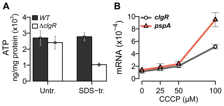
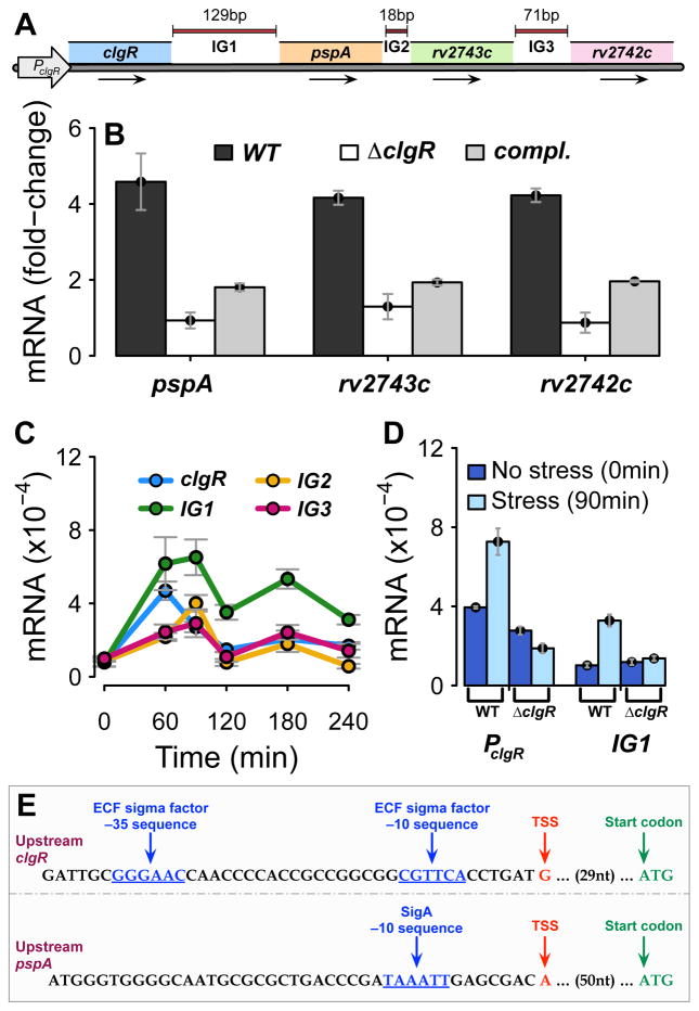
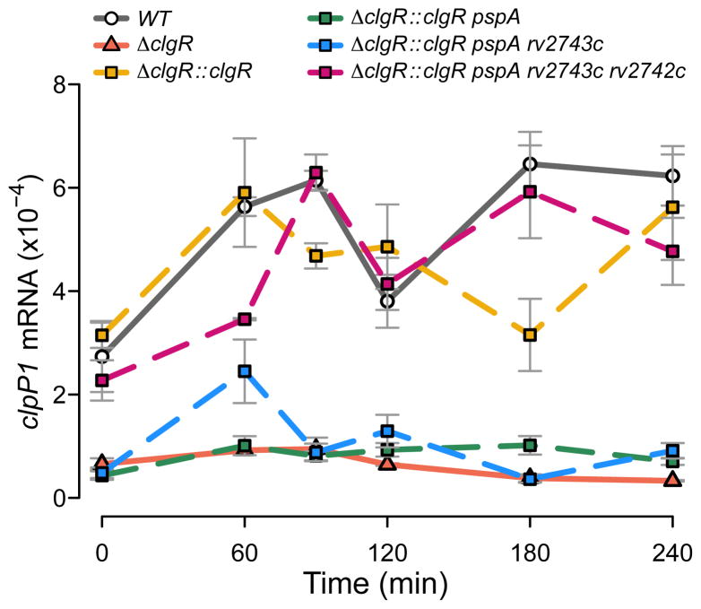
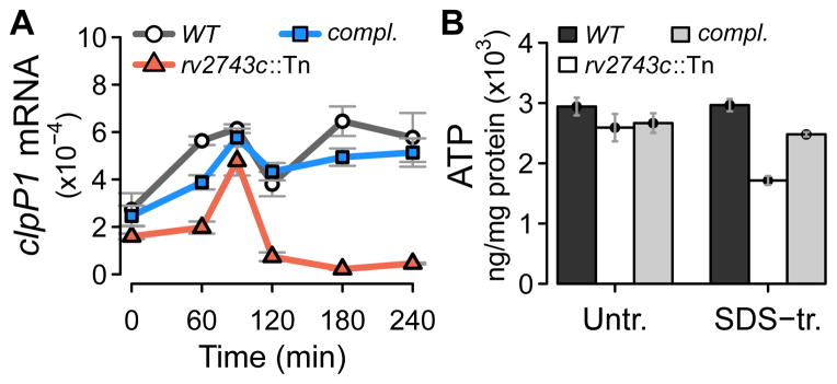
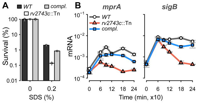
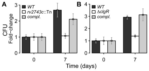
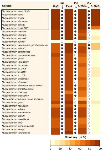
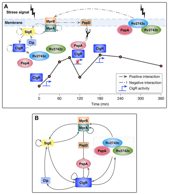
The bacterial envelope integrates essential stress-sensing and adaptive functions; thus, envelope-preserving functions are important for survival. In Gram-negative bacteria, envelope integrity during stress is maintained by the multi-gene Psp response. Mycobacterium tuberculosis was thought to lack the Psp system since it encodes only pspA and no other psp ortholog. Intriguingly, pspA maps downstream from clgR, which encodes a transcription factor regulated by the MprAB-σ(E) envelope-stress-signaling system. clgR inactivation lowered ATP concentration during stress and protonophore treatment-induced clgR-pspA expression, suggesting that these genes express Psp-like functions. We identified a four-gene set - clgR, pspA (rv2744c), rv2743c, rv2742c - that is regulated by clgR and in turn regulates ClgR activity. Regulatory and protein-protein interactions within the set and a requirement of the four genes for functions associated with envelope integrity and surface-stress tolerance indicate that a Psp-like system has evolved in mycobacteria. Among Actinobacteria, the four-gene module occurred only in tuberculous mycobacteria and was required for intramacrophage growth, suggesting links between its function and mycobacterial virulence. Additionally, the four-gene module was required for MprAB-σ(E) stress-signaling activity. The positive feedback between envelope-stress-sensing and envelope-preserving functions allows sustained responses to multiple, envelope-perturbing signals during chronic infection, making the system uniquely suited to tuberculosis pathogenesis.


Conflict of interest statement

The authors have no conflict of interest to declare.

Figures

Figure 1
Figure 1
(A) Effect of clgR deletion on intracellular ATP levels. Mid-log cultures of wild-type and clgR mutant strains were treated with 0.03% SDS for 24 hrs. Extracts from pre- and post-treatment cultures were then assayed for ATP levels and expressed as ng of ATP per mg of total protein. (B) Effect of treatment with carbonyl cyanide m-chlorophenyl hydrazone (CCCP) on expression of clgR and pspA. Mid-log cultures of the wild-type strain were treated for 6 hrs with various concentrations of CCCP, as indicated, and harvested pre- and post-treatment. Total RNA was extracted, and amplicons were measured by quantitative RT-PCR using primers and gene-specific molecular beacons. Amplicon copy numbers were normalized to 16S rRNA. 0 μM = solvent-only control (corresponding to the highest solvent concentration used with the CCCP treatment). In this and subsequent figures, quantitative RT-PCR data are presented as mean values (+/− standard error of the mean) from triplicate experiments.
Figure 2
Figure 2. Structure and regulation of genes in the clgR-pspA-rv2743c-rv2742c region
(A) Schematic representation of the region. Arrows represent direction of transcription. The arrow PclgR indicates sequences upstream of clgR expressing promoter activity. IG = intergenic regions of indicated length, in bp. (B) Effect of clgR deletion on gene stress response. Mid-log cultures of wild-type, clgR deletion mutant, and complemented strains were treated with 0.03% SDS and harvested pre-treatment (time 0) and post-treatment (90 min). Amplicons were generated from total RNA by quantitative RT-PCR using primers and gene-specific molecular beacons from intragenic sequences for each gene, as indicated. Amplicon copy numbers, normalized to 16S rRNA, are presented as fold-change relative to time 0. Mean values (+/− standard error of the mean) were obtained from triplicate experiments. (C) Enumeration of intergenic-sequence-containing amplicons. Mid-log cultures of the wild-type strain were treated with SDS, and samples were harvested pre-treatment and at multiple times post treatment. Amplicons were generated from sequences internal to clgR (reference gene) and from intergenic sequences along the region, as indicated (see panel A). Data obtained from triplicate experiments were normalized to 16S rRNA. (Note that copy numbers of RT-PCR amplicons obtained with different primer sets and molecular beacons cannot be compared with each other due to variations in primer-associated efficiency of PCR amplification and molecular-beacon-dependent detection sensitivity). (D) Analysis of promoter probe constructs. Two promoter::lacZ fusions were generated with nucleotide sequences comprising either 382 bp upstream of clgR (PclgR, see panel A) or 250 bp upstream of pspA (IG1, see panel A) in wild-type and clgR mutant strains. lacZ transcripts were enumerated by quantitative RT-PCR prior to and 90 min following SDS treatment of mid-log cultures. Data obtained from triplicate experiments were normalized to 16S rRNA. (E) Promoter mapping upstream of clgR and pspA. The panel shows nucleotide sequences upstream of clgR and upstream of pspA (intergenic region IG1). Indicated in brown are the two transcription start sites (TSS) mapped in this work using the genome-wide mapping method described in Methods. The TSS in the top row had a ratio of converted to unconverted 5′ ends >1.74 (adjusted p ≤0.01 for probability of being a processed 5′ end). This TSS is preceded by −10 and −35 sequences (shown in blue) characteristic of the extracytoplasmic function (ECF) sigma factor SigE (Manganelli et al., 2001). The 5′ end in the bottom row had a converted/non-converted ratio of 1.58, and was confirmed to be a TSS based on the presence of a very strong SigA −10 motif (in blue) (Cortes et al., 2013, Feklistov & Darst, 2011). In both rows, the annotated first codon (ATG) is as reported in the M. tuberculosis genome sequence (http://tuberculist.epfl.ch/). nt = nucleotides.
Figure 3
Figure 3. Effect on clpP1 stress response of clgR deletion and sequential complementation with clgR and downstream operon genes
Expression of the sentinel target gene clpP1 was used as readout of ClgR activity (Sherrid et al., 2010). Complementation of the clgR deletion was performed sequentially by introducing clgR alone, clgR plus pspA, plus pspA-rv2743c, and plus pspA-rv2743c-rv2742c. Transcript copy numbers were generated and expressed as described in the legend to Fig. 2C. The reason why clgR alone complements the clpP1 expression defect of the clgR deletion mutant to similar levels as the clgR-pspA-rv2743c-rv2742c complementation can be explained by ectopically produced ClgR inducing pspA-rv2743c-rv2742c expression from the promoter located in the clgR-pspA intergenic region (Fig. 2D). Measurements of clpP1 transcripts performed using M. tuberculosis H37Rv and M. tuberculosis CDC1551 gave indistinguishable results (Fig. S8D), indicating that the clpP1 surface-stress response is conserved in different M. tuberculosis strains.
Figure 4
Figure 4. Effects of rv2743c inactivation on target gene expression and ATP levels
(A) Effect on clpP1 stress response. ClgR activity was assessed as expression of the sentinel target gene clpP1 in an rv2743c transposon (Tn) insertion mutant. Complementation was performed with rv2743c-rv2742c to overcome the polar effect of the mutation on the downstream rv2742c (data not shown). Wild-type, mutant, and rv2743c-rv2742c complemented (compl.) strains were subjected to surface stress with SDS, and clpP1 transcripts were enumerated and presented as described in the legend to Fig. 2C. The reason why the rv2743c mutation did not phenocopy the clgR-pspA complementation of the clgR deletion mutant (Fig. 3) can be gene dosage effect due to the extra copy of pspA in the complementing DNA. (B) Effect on intracellular ATP levels. ATP levels were assayed in extracts from mid-log cultures of wild-type, mutant, and complemented strains pre- and post-treatment with 0.03% SDS for 24 hrs, and expressed as in the legend to Fig. 1A.
Figure 5
Figure 5. Protein-protein interactions
Pairwise protein-protein interactions were assessed between differentially tagged recombinant proteins expressed from IPTG-inducible promoters in E. coli. Whole cell lysates from IPTG-induced recombinant cultures expressing tagged proteins, alone or in pairs as indicated, were used for co-immunoprecipitation (IP; left column) or for pull-down of 6xHis tagged protein (PD; right column), followed by immunoblotting (IB) with the appropriate monoclonal antibody. Left column: IP was performed with anti-His (A and C) or with anti-S-tag antibody (B), followed by IB with anti-Myc antibody to detect ClgR. Lanes: 1) whole-cell lysate + IB (positive control); 2) IP (with cell extracts carrying ClgR-Myc alone) + IB; 3) IP (with cell extracts carrying ClgR-Myc plus a His-tagged or S-tagged protein) + IB. Right column: PD was performed with Ni-NTA agarose, followed by IB with anti S-tag antibody for detection of Rv2743c (D and E) or Rv2742c (F). Lanes: 1) whole-cell lysate + IB (positive control); 2) PD (with cell extracts carrying S-tag protein alone) + IB; 3) PD (with cell extracts carrying S-tag protein plus His-tag protein) + IB. The bottom row of each panel shows unidentified E. coli bands cross-reacting with anti-Myc monoclonal antibody (panels in the left column) or with the anti-S-tag monoclonal antibody (panels in the right column) that served as loading controls. Experiments in panels B, C, and F were repeated by switching tags in each corresponding protein pair to control for potential tag effects on the protein-protein interaction, with no change of result. S = S tag; M = Myc tag; H = His tag.
Figure 6
Figure 6. Effects of rv2743c inactivation
(A) Tolerance to surface stress. Lethal activity of the detergent SDS was determined in wild-type, rv2743c mutant and rv2743c-rv2742c complemented (compl.) strains. Data are expressed as % survival relative to untreated cultures. Mean values (+/− standard error of the mean) from triplicate experiments are shown. (B) Surface-stress response of sentinel target genes of MprAB and σE. Transcripts for mprA and sigB were enumerated in wild-type, mutant, and complemented strains and presented as described in the legend to Fig. 2C. Tn = transposon.
Figure 7
Figure 7. Effect of rv2743c and clgR inactivation for growth in macrophages
Human monocyte-derived macrophages were infected with different M. tuberculosis strains. Bacterial counts (CFU) were normalized to 105 adherent cells per well. Data are expressed as fold-increase in CFU at day 7 relative to CFU enumerated at 4 hrs post-infection. Mean values (+/− standard error of the mean) from triplicate experiments are shown. (A) Wild-type, rv2743c mutant and rv2743c-rv2742c complemented (compl.) strains. Tn = transposon. (B) Wild-type, clgR mutant and complemented (compl.) strains. Complementation was with clgR-pspA-rv2743c-rv2742c DNA. Reduced growth of the clgR mutant in murine bone-marrow-derived macrophages was previously reported (Estorninho et al., 2010).
Figure 8
Figure 8. M. tuberculosis (Mtb) and E. coli (Eco) Psp protein comparison
(A) Mtb ClgR and Eco PspF. ClgR and PspF contain helix-turn-helix (HTH)-type DNA-binding domains (DBD). ClgR has an N-terminal HTH_XRE (SM00530, PF01381) DBD, while PspF has a C-terminal Fis-type HTH_8 DBD (PF02954, IPR002197). PspF contains AAA+ and sigma-54 interaction domains (IPR003593 and IPR002078, respectively). (B) Mtb PspA and Eco PspA. Both proteins were used to define PspA/IM30 (PF04012, IPR007157), a domain characterized by ~25% of conserved amino acid residues spanning the entire protein length. This domain is also predicted to contain coiled-coils. (C) Mtb Rv2743c and Eco PspB. Mtb Rv2743c and Eco PspB do not share significant sequence similarity; however, they are both predicted to be transmembrane (TM) proteins. Mtb Rv2743c is predicted to have two ~22-amino-acid TM helices connected by a short (4–6 amino acid) non-cytoplasmic loop. Mtb Rv2743c is also predicted to contain a C-terminal coiled-coil. Eco PspB is a single PspB domain (PF06667, IPR009554), which contains a predicted N-terminal TM helix (residues: 6–24) and C-terminal coiled-coil (residues: 36–64). (D) Mtb Rv2742c and Eco PspC. Mtb Rv2742c and Eco PspC share no significant sequence similarity. Sequence analysis of Rv2742c revealed only a single domain of unknown function (DUF3046, PF11248), which is conserved in actinobacteria. In contrast, Eco PspC has an N-terminal PspC domain (residues: 7–68, PF04024), a transmembrane helix (residues: 39–64), and a C-terminal coiled-coil (residues: 84–105).
Figure 9
Figure 9. Distribution of ClgR-PspA-Rv2743c-Rv2742c proteins in Mycobacterium spp
Significant matches and similarity scores were obtained as described in Methods. The heat map represents the similarity score per target species or sub-species (or the highest score in the case of multiple matches), expressed as percent of protein sequence similarity for the longest contiguous match. Mycobacterial species were ordered on the basis of phylogenetic similarity ((Wattam et al., 2014) and http://www.patricbrc.org). The box contains the species in the M. tuberculosis complex. Intergenic distances (IG) between matches were calculated (in bp) in the target genomes. IG1, IG2 and IG3 denote distances between clgR-pspA, pspA-rv2743c and rv2743c-rv2742c, respectively. Filled black circles indicate intergenic distances that are similar to that of M. tuberculosis clgR-pspA-rv2743c-rv2742c operon, as shown in Fig. 2A. Open circles denote either absence of one gene in the pair, intergenic distances of >250 bp, or absence of genomic location information. *Of 13 virulent M. bovis isolates analyzed, one (AF2122/97) contained a 23-bp insertion in pspC approximately two-thirds into the gene sequence, presumably giving rise to a truncated product. The same insertion was found in the genomes of 9 out of 14 substrains of the attenuated vaccine strain M. bovis Bacillus Calmette Guerin. The evolutionary significance of this polymorphism remains to be ascertained. **Only Mycobacterium leprae was devoid of the entire ClgR-Psp module, as previously reported (Bretl et al., 2014), suggesting that loss of the Psp system is part of the genomic downsizing observed with this obligate intracellular pathogen (Cole et al., 2001). ***Only Mycobacterium avium among non-tuberculous mycobacteria showed a truncated form of Rv2742c (~1/3 the length of M. tuberculosis Rv2742c) at the appropriate distance from the preceding gene.
Figure 10
Figure 10
(A) Model of MprAB-σE-dependent maintenance of envelope integrity in response to surface stress. The figure shows interactions between the envelope-stress signaling MprAB-σE network and the envelope-preserving ClgR-PspA-Rv2743c-Rv2742c system. Interactions include the mprAB-sigE transcriptional network and the mprAB-dependent pepD, the downstream target clgR-pspA-rv2743c-rv2742c operon, and additional clgR-regulated genes (clpP1). The line plot represents the temporal, post-stress expression profile of clpP1, as a proxy of ClgR activity. The location of individual proteins, protein complexes, and interactions relative to the line plot depicts the proposed temporal course of events associated with propagation of signal (ClgR activity) through the Psp system, resulting membrane integrity, and MprAB stabilization. The scissors icon indicates proteolytic degradation. The red cross over the ClgR activity icon represents ClgR inactivation. (B) Feedback loops in the stress-response system of M. tuberculosis. The wiring diagram shows multiple regulatory feedback loops, transcriptional and post-transcriptional, operating between mprAB, sigE, clgR, the Psp system, and neighboring network genes and/or corresponding gene products under surface stress conditions. The abundance of feedback loops in the system does not allow distinguishing whether the observed functional effects of the pspA-rv2743c-rv2742c products are direct, or occur indirectly via the regulation of ClgR and its target gene products. Since clgR is connected to the mprAB/sigE sensing network through two positive feedbacks, links between envelope-stress-sensing and envelope-preserving functions exist regardless of whether pspA-rv2743c-rv2742c act directly or indirectly. Except for sigE autoregulation (Chauhan and Gennaro, unpublished), the regulatory interactions showed in this figure are all cited or discovered in the present manuscript. Arrowhead = positive regulation; Barhead = negative regulation.

References

    1. Alber T. Signaling mechanisms of the Mycobacterium tuberculosis receptor Ser/Thr protein kinases. Curr Opin Struct Biol. 2009;19:650–657. - PMC - PubMed
    1. Altschul SF, Gish W, Miller W, Myers EW, Lipman DJ. Basic local alignment search tool. J Mol Biol. 1990;215:403–410. - PubMed
    1. Altschul SF, Madden TL, Schaffer AA, Zhang J, Zhang Z, Miller W, Lipman DJ. Gapped BLAST and PSI-BLAST: a new generation of protein database search programs. Nucleic Acids Res. 1997;25:3389–3402. - PMC - PubMed
    1. Barik S, Sureka K, Mukherjee P, Basu J, Kundu M. RseA, the SigE specific anti-sigma factor of Mycobacterium tuberculosis, is inactivated by phosphorylation-dependent ClpC1P2 proteolysis. Mol Microbiol. 2010;75:592–606. - PubMed
    1. Becker LA, Bang IS, Crouch ML, Fang FC. Compensatory role of PspA, a member of the phage shock protein operon, in rpoE mutant Salmonella enterica serovar Typhimurium. Mol Microbiol. 2005;56:1004–1016. - PubMed

Publication types

MeSH terms