

Blood pressure and sodium: Association with MRI markers in cerebral small vessel disease
- PMID: 25899292
- PMCID: PMC4758556
- DOI: 10.1038/jcbfm.2015.64
Blood pressure and sodium: Association with MRI markers in cerebral small vessel disease
Abstract
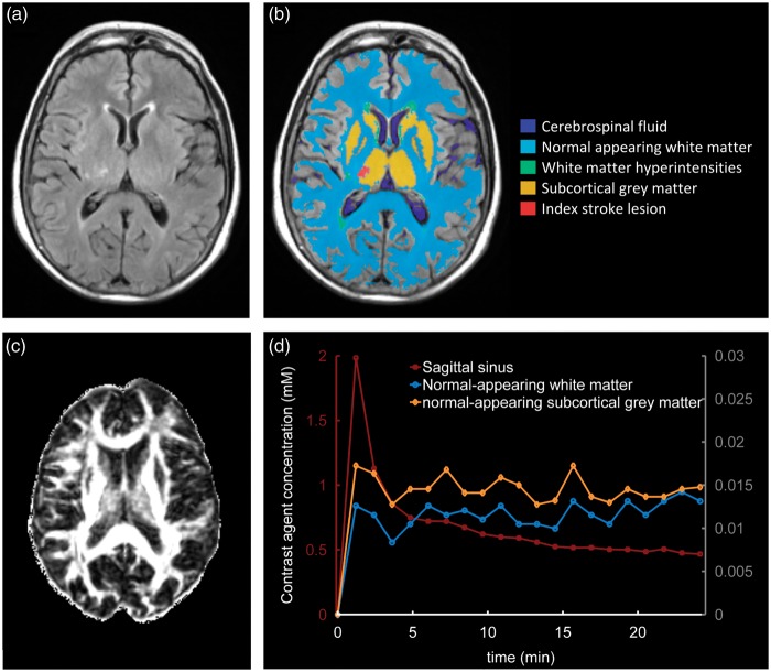
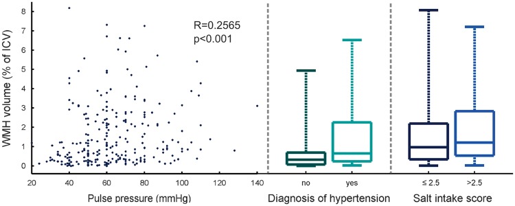
Dietary salt intake and hypertension are associated with increased risk of cardiovascular disease including stroke. We aimed to explore the influence of these factors, together with plasma sodium concentration, in cerebral small vessel disease (SVD). In all, 264 patients with nondisabling cortical or lacunar stroke were recruited. Patients were questioned about their salt intake and plasma sodium concentration was measured; brain tissue volume and white-matter hyperintensity (WMH) load were measured using structural magnetic resonance imaging (MRI) while diffusion tensor MRI and dynamic contrast-enhanced MRI were acquired to assess underlying tissue integrity. An index of added salt intake (P = 0.021), pulse pressure (P = 0.036), and diagnosis of hypertension (P = 0.0093) were positively associated with increased WMH, while plasma sodium concentration was associated with brain volume (P = 0.019) but not with WMH volume. These results are consistent with previous findings that raised blood pressure is associated with WMH burden and raise the possibility of an independent role for dietary salt in the development of cerebral SVD.
Figures
References
-
- Maclullich AM, Ferguson KJ, Reid LM, Deary IJ, Starr JM, Seckl JR, et al. Higher systolic blood pressure is associated with increased water diffusivity in normal-appearing white matter. Stroke 2009; 40: 3869–3871. - PubMed
-
- Decaux G. Is asymptomatic hyponatremia really asymptomatic? Am J Med 2006; 119: S79–S82. - PubMed
Publication types
MeSH terms
Substances
Grants and funding
LinkOut - more resources
Full Text Sources
Other Literature Sources
Medical
