

Longitudinal change of small-vessel disease-related brain abnormalities
- PMID: 25899293
- PMCID: PMC4758559
- DOI: 10.1038/jcbfm.2015.72
Longitudinal change of small-vessel disease-related brain abnormalities
Abstract
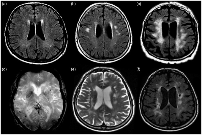
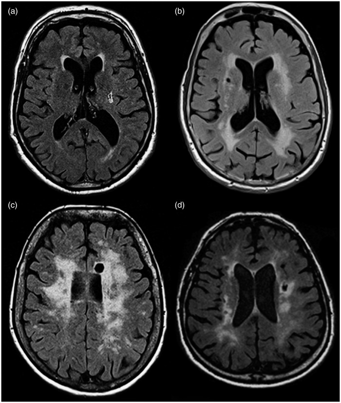
Knowledge about the longitudinal change of cerebral small-vessel disease–related magnetic resonance imaging abnormalities increases our pathophysiologic understanding of cerebral microangiopathy. The change of specific lesion types may also serve as secondary surrogate endpoint in clinical trials. A surrogate endpoint needs to progress fast enough to allow monitoring of treatment effects within a reasonable time period, and change of the brain abnormality needs to be correlated with clinical change. Confluent white matter lesions show fast progression and correlations with cognitive decline. Thus, the change of confluent white matter lesions may be used as a surrogate marker in proof-of-concept trials with small patient numbers needed to show treatment effects on lesion progression. Nonetheless if the expected change in cognitive performance resulting from treatment effects on lesion progression is used as outcome, the sample size needed to show small to moderate treatment effects becomes very large. Lacunes may also fulfill the prerequisites of a surrogate marker, but in the general population the incidence of lacunes over short observational periods is small. For other small-vessel disease–related brain abnormalities including microbleeds and microstructural changes in normal-appearing white matter longitudinal change and correlations with clinical decline is not yet fully determined.
Figures
References
-
- Pantoni L. Cerebral small vessel disease: from pathogenesis and clinical characteristics to therapeutic challenges. Lancet Neurol 2010; 9: 689–701. - PubMed
-
- Schmidt R, Schmidt H, Haybaeck J, Loitfelder M, Weis S, Cavalieri M, et al. Heterogeneity in age-related white matter changes. Acta Neuropathol 2011; 122: 171–185. - PubMed
-
- Cavalieri M, Schmidt H, Schmidt R. Structural MRI in normal aging and Alzheimer’s disease: white and black spots. Neurodegener Dis 2012; 10: 253–256. - PubMed
-
- Poggesi A, Pantoni L, Inzitari D, Fazekas F, Ferro J, O'Brien J, et al. The LADIS Study Group. 2001–2011: a decade of the LADIS (Leukoaraiosis And DISability) Study: what have we learned about white matter changes and small-vessel disease? Cerebrovasc Dis 2011; 32: 577–588. - PubMed
-
- Graham SJ, Henkelman RM. Understanding pulsed magnetization transfer. J Magn Reson Imaging 1997; 7: 903–912. - PubMed
Publication types
MeSH terms
LinkOut - more resources
Full Text Sources
Other Literature Sources
