

Cerebral perfusion and glucose metabolism in Alzheimer's disease and frontotemporal dementia: two sides of the same coin?
- PMID: 25899416
- PMCID: PMC4562004
- DOI: 10.1007/s00330-015-3696-1
Cerebral perfusion and glucose metabolism in Alzheimer's disease and frontotemporal dementia: two sides of the same coin?
Abstract
Objectives: Alzheimer's disease (AD) and frontotemporal (FTD) dementia can be differentiated using [(18)F]-2-deoxy-2-fluoro-D-glucose (FDG)-PET. Since cerebral blood flow (CBF) is related to glucose metabolism, our aim was to investigate the extent of overlap of abnormalities between AD and FTD.
Methods: Normalized FDG-PET and arterial spin labelling (ASL-MRI)-derived CBF was measured in 18 AD patients (age, 64 ± 8), 12 FTD patients (age, 61 ± 8), and 10 controls (age, 56 ± 10). Voxel-wise comparisons, region-of-interest (ROI), correlation, and ROC curve analyses were performed.
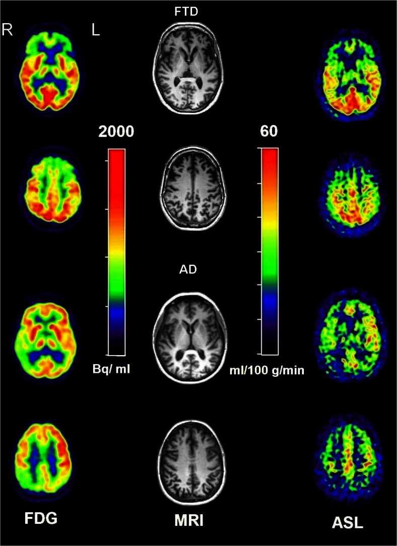
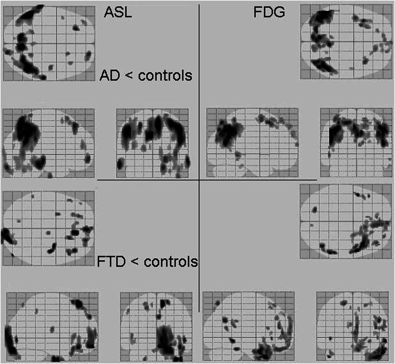
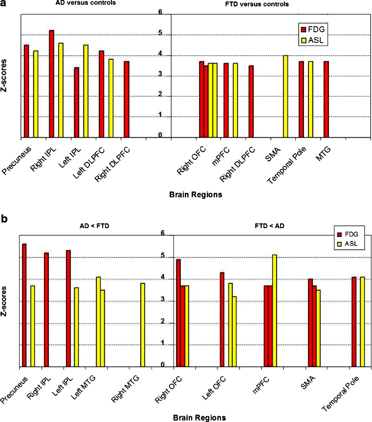
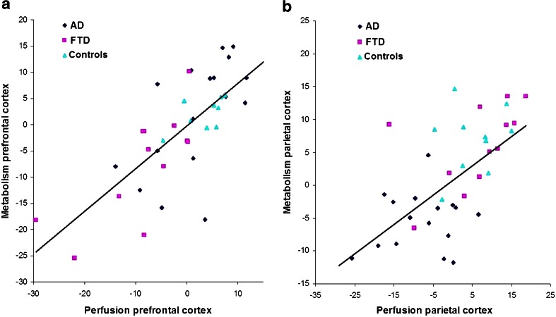
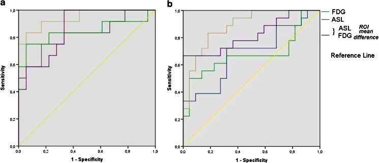
Results: Voxel-wise comparisons showed decreased CBF and FDG uptake in AD compared with controls and FTD in both precuneus and inferior parietal lobule (IPL). Compared with controls and AD, FTD patients showed both hypometabolism and hypoperfusion in medial prefrontal cortex (mPFC). ASL and FDG were related in precuneus (r = 0.62, p < 0.001), IPL (r = 0.61, p < 0.001), and mPFC across groups (r = 0.74, p < 001). ROC analyses indicated comparable performance of perfusion and metabolism in the precuneus (AUC, 0.72 and 0.74), IPL (0.85 and 0.94) for AD relative to FTD, and in the mPFC in FTD relative to AD (both 0.68).
Conclusions: Similar patterns of hypoperfusion and hypometabolism were observed in regions typically associated with AD and FTD, suggesting that ASL-MRI provides information comparable to FDG-PET.
Key points: • Similar patterns of hypoperfusion and hypometabolism were observed in patients with dementia. • For both imaging modalities, parietal abnormalities were found in Alzheimer's disease. • For both imaging modalities, prefrontal abnormalities were found in frontotemporal dementia.
Figures
Similar articles
-
Alzheimer Disease and Mild Cognitive Impairment: Integrated Pulsed Arterial Spin-Labeling MRI and 18F-FDG PET.Radiology. 2018 Jul;288(1):198-206. doi: 10.1148/radiol.2018170575. Epub 2018 May 15. Radiology. 2018. PMID: 29762090
-
Arterial spin labeling versus 18F-FDG-PET to identify mild cognitive impairment.Neuroimage Clin. 2020;25:102146. doi: 10.1016/j.nicl.2019.102146. Epub 2019 Dec 23. Neuroimage Clin. 2020. PMID: 31931403 Free PMC article.
-
Using simultaneous PET/MRI to compare the accuracy of diagnosing frontotemporal dementia by arterial spin labelling MRI and FDG-PET.Neuroimage Clin. 2017 Oct 31;17:405-414. doi: 10.1016/j.nicl.2017.10.033. eCollection 2018. Neuroimage Clin. 2017. PMID: 29159053 Free PMC article.
-
Brain fluorodeoxyglucose (FDG) PET in dementia.Ageing Res Rev. 2016 Sep;30:73-84. doi: 10.1016/j.arr.2016.02.003. Epub 2016 Feb 11. Ageing Res Rev. 2016. PMID: 26876244 Review.
-
Positron emission tomography imaging in dementia.Br J Radiol. 2007 Dec;80 Spec No 2:S160-7. doi: 10.1259/bjr/97295129. Br J Radiol. 2007. PMID: 18445746 Review.
Cited by
-
Neuroimaging in genetic frontotemporal dementia and amyotrophic lateral sclerosis.Neurobiol Dis. 2020 Nov;145:105063. doi: 10.1016/j.nbd.2020.105063. Epub 2020 Sep 2. Neurobiol Dis. 2020. PMID: 32890771 Free PMC article. Review.
-
Imaging and fluid biomarkers in frontotemporal dementia.Nat Rev Neurol. 2017 Jul;13(7):406-419. doi: 10.1038/nrneurol.2017.75. Epub 2017 Jun 16. Nat Rev Neurol. 2017. PMID: 28621768 Review.
-
Sympathetic Innervation of Cold-Activated Brown and White Fat in Lean Young Adults.J Nucl Med. 2017 May;58(5):799-806. doi: 10.2967/jnumed.116.180992. Epub 2016 Oct 27. J Nucl Med. 2017. PMID: 27789721 Free PMC article.
-
Diagnostic imaging of dementia with Lewy bodies, frontotemporal lobar degeneration, and normal pressure hydrocephalus.Jpn J Radiol. 2020 Jan;38(1):64-76. doi: 10.1007/s11604-019-00881-9. Epub 2019 Sep 23. Jpn J Radiol. 2020. PMID: 31549279 Review.
-
Estimating regional cerebral blood flow using resting-state functional MRI via machine learning.J Neurosci Methods. 2020 Feb 1;331:108528. doi: 10.1016/j.jneumeth.2019.108528. Epub 2019 Nov 19. J Neurosci Methods. 2020. PMID: 31756399 Free PMC article.
References
Publication types
MeSH terms
Substances
LinkOut - more resources
Full Text Sources
Other Literature Sources
Medical
