Review
. 2015 Jun 18;125(25):3851-9.
doi: 10.1182/blood-2015-01-544676. Epub 2015 Apr 21.

Regulation of thrombosis and vascular function by protein methionine oxidation

Affiliations
Review

Regulation of thrombosis and vascular function by protein methionine oxidation

Sean X Gu et al. Blood. .

Abstract

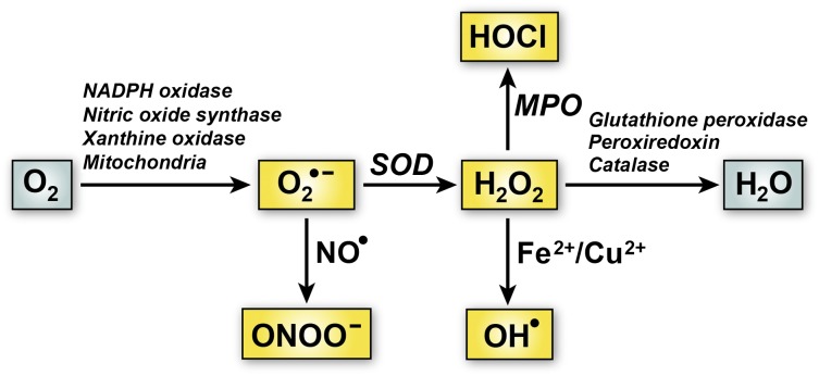
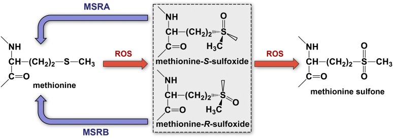
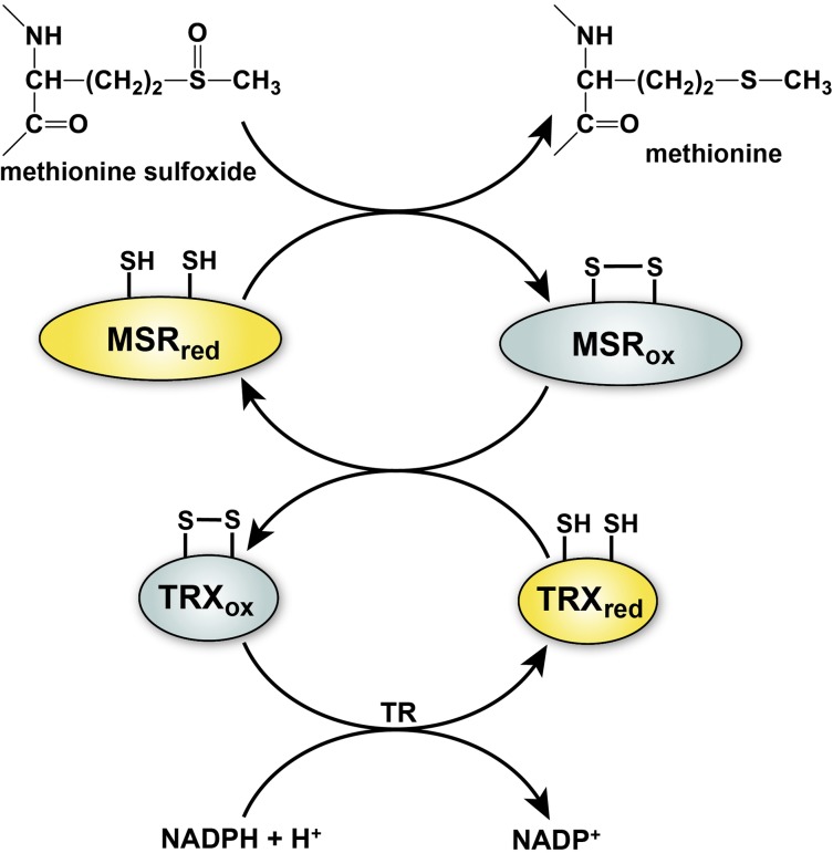
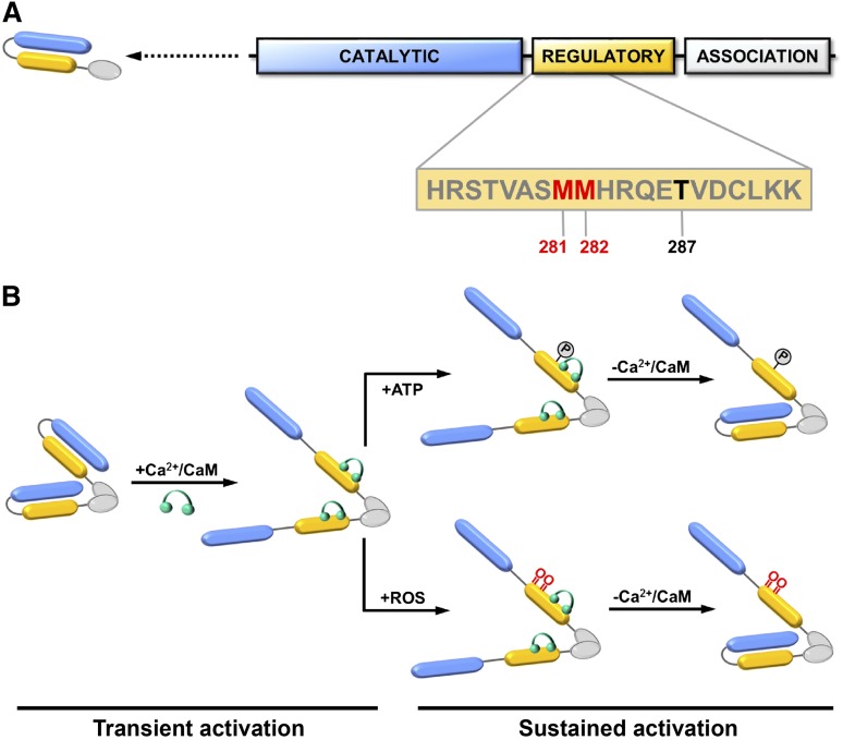
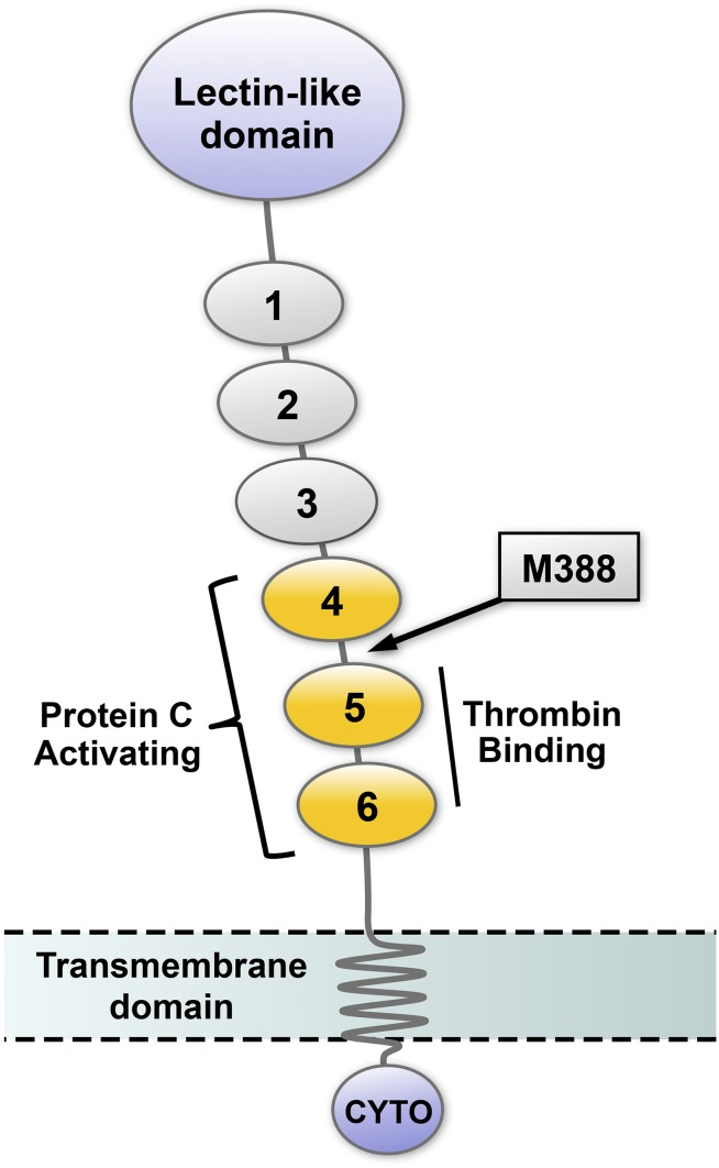
Redox biology is fundamental to both normal cellular homeostasis and pathological states associated with excessive oxidative stress. Reactive oxygen species function not only as signaling molecules but also as redox regulators of protein function. In the vascular system, redox reactions help regulate key physiologic responses such as cell adhesion, vasoconstriction, platelet aggregation, angiogenesis, inflammatory gene expression, and apoptosis. During pathologic states, altered redox balance can cause vascular cell dysfunction and affect the equilibrium between procoagulant and anticoagulant systems, contributing to thrombotic vascular disease. This review focuses on the emerging role of a specific reversible redox reaction, protein methionine oxidation, in vascular disease and thrombosis. A growing number of cardiovascular and hemostatic proteins are recognized to undergo reversible methionine oxidation, in which methionine residues are posttranslationally oxidized to methionine sulfoxide. Protein methionine oxidation can be reversed by the action of stereospecific enzymes known as methionine sulfoxide reductases. Calcium/calmodulin-dependent protein kinase II is a prototypical methionine redox sensor that responds to changes in the intracellular redox state via reversible oxidation of tandem methionine residues in its regulatory domain. Several other proteins with oxidation-sensitive methionine residues, including apolipoprotein A-I, thrombomodulin, and von Willebrand factor, may contribute to vascular disease and thrombosis.


Figures

Figure 1
Figure 1
Major pathways of ROS generation and elimination in vascular cells. One electron reduction of O2 can produce superoxide anion (O2•−). The main pathways for O2 transformation are via dismutation to hydrogen peroxide (H2O2) catalyzed by superoxide dismutase (SOD) or reaction with nitric oxide (NO) to form peroxynitrite (ONOO). H2O2 can be further metabolized to hypochlorous acid (HOCl) by myeloperoxidase (MPO) or hydroxyl radical (OH) through reactions catalyzed by metal ions (Fe2+ or Cu2+) via the Fenton reaction. Elimination of H2O2 is facilitated by antioxidant enzymes such as glutathione peroxidase, peroxiredoxin, and catalase.
Figure 2
Figure 2
Biochemistry of protein methionine oxidation and reduction. Oxidation of protein methionine residues produces 2 sulfoxide diastereomers, methionine-S-sulfoxide and methionine-R-sulfoxide, which can be stereospecifically reduced back to methionine by 2 classes of mammalian methionine sulfoxide reductases, MSRA and MSRB, respectively. Further oxidation of methionine sulfoxide to methionine sulfone is biologically irreversible.
Figure 3
Figure 3
General mechanism of MSR and regeneration by the thioredoxin system. Reduction of methionine sulfoxide to methionine by MSR results in the transient formation of an intramolecular disulfide bond that inactivates the enzyme. Disulfide exchange reaction with thioredoxin (TRX) regenerates the active form of MSR and leads to inactivation of TRX. Regeneration of reduced TRX occurs through a transfer of electrons from NADPH in a reaction catalyzed by thioredoxin reductase (TR). The active reduced forms of the enzymes are depicted in yellow and the inactive oxidized forms of enzymes in gray.
Figure 4
Figure 4
CaMKII domain structure and activation. (A) Each CaMKII monomer contains a catalytic kinase domain (blue), a regulatory domain involved (yellow), and an association domain (white). Dimerization of monomeric subunits is mediated via the association domains, followed by oligomerization to form a holoenzyme consisting of 2 stacked hexameric rings (not shown). The sequence of the regulatory domain containing 2 redox-active methionine residues (Met281/Met282) (red) and Thr287 (black) are indicated. (B) In the resting state, the kinase activity of CaMKII is autoinhibited by an interaction between its catalytic and regulatory domains. Binding of calcium/calmodulin (Ca2+/CaM) (green) to the regulatory domain causes disassociation and transient activation of the catalytic domain. Autophosphorylation at Thr-287 or oxidation at Met281/Met282 prevents autoinhibition and causes sustained activation of CaMKII that is independent of calcium/calmodulin binding. Adapted with permission from Scott et al.
Figure 5
Figure 5
Schematic representation of thrombomodulin domain structure. The amino terminal portion of TM, which projects into the vascular lumen, contains a lectin-like domain and 6 EGF-like domains. EGF-like domains 5 and 6 are required for thrombin binding, and EGF-like domains 4 to 6 (yellow) are necessary for efficient activation of protein C. The location of the oxidation-sensitive regulatory methionine (M388) is in the short linker region between EGF-like domains 4 and 5 (arrow). TM also contains a transmembrane domain and a cytoplasmic tail (CYTO).

References

    1. Taniyama Y, Griendling KK. Reactive oxygen species in the vasculature: molecular and cellular mechanisms. Hypertension. 2003;42(6):1075–1081. - PubMed
    1. Kim YW, Byzova TV. Oxidative stress in angiogenesis and vascular disease. Blood. 2014;123(5):625–631. - PMC - PubMed
    1. Dikalov SI, Harrison DG. Methods for detection of mitochondrial and cellular reactive oxygen species. Antioxid Redox Signal. 2014;20(2):372–382. - PMC - PubMed
    1. Sugamura K, Keaney JF., Jr Reactive oxygen species in cardiovascular disease. Free Radic Biol Med. 2011;51(5):978–992. - PMC - PubMed
    1. Sesso HD, Buring JE, Christen WG, et al. Vitamins E and C in the prevention of cardiovascular disease in men: the Physicians’ Health Study II randomized controlled trial. JAMA. 2008;300(18):2123–2133. - PMC - PubMed

Publication types