. 2015 Jul;14(7):1781-95.
doi: 10.1074/mcp.M114.045658. Epub 2015 Apr 21.

BioID-based Identification of Skp Cullin F-box (SCF)β-TrCP1/2 E3 Ligase Substrates

Affiliations

BioID-based Identification of Skp Cullin F-box (SCF)β-TrCP1/2 E3 Ligase Substrates

Etienne Coyaud et al. Mol Cell Proteomics. 2015 Jul.

Abstract

The identification of ubiquitin E3 ligase substrates has been challenging, due in part to low-affinity, transient interactions, the rapid degradation of targets and the inability to identify proteins from poorly soluble cellular compartments. SCF(β-TrCP1) and SCF(β-TrCP2) are well-studied ubiquitin E3 ligases that target substrates for proteasomal degradation, and play important roles in Wnt, Hippo, and NFκB signaling. Combining 26S proteasome inhibitor (MG132) treatment with proximity-dependent biotin labeling (BioID) and semiquantitative mass spectrometry, here we identify SCF(β-TrCP1/2) interacting partners. Based on their enrichment in the presence of MG132, our data identify over 50 new putative SCF(β-TrCP1/2) substrates. We validate 12 of these new substrates and reveal previously unsuspected roles for β-TrCP in the maintenance of nuclear membrane integrity, processing (P)-body turnover and translational control. Together, our data suggest that β-TrCP is an important hub in the cellular stress response. The technique presented here represents a complementary approach to more standard IP-MS methods and should be broadly applicable for the identification of substrates for many ubiquitin E3 ligases.


Figures

Fig. 1.
Fig. 1.
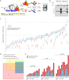
BioID can be used to identify ubiquitin E3 ligase substrates. A, An N-terminal tag consisting of the FLAG epitope and the mutant E. coli biotin conjugating protein BirA R118G (BirA*) was fused to the N terminus of the human F-box proteins β-TrCP1 and β-TrCP2. The BirA* protein converts biotin (black hexagon) to biotinoyl-AMP (yellow hexagon). The mutant BirA protein exhibits a reduced affinity for the activated biotin molecule; biotinoyl-AMP thus diffuses away and reacts with free amine groups on lysine residues in nearby polypeptides, including e.g. the bait protein itself, other SCF complex components (CUL1, SKP1), SCF substrates (S) and substrate binding partners (A, B). In the presence of the 26S proteasome inhibitor MG132, β-TrCP substrates are stabilized. Following cell lysis under stringent buffer conditions, biotinylated proteins are affinity purified using streptavidin coupled to Sepharose beads. Streptavidin-bound proteins are washed and subjected to trypsin proteolysis, and the liberated peptides are identified using tandem mass spectrometry. B, Expression of FLAGBirA*-β-TrCP1/2 leads to biotinylation of endogenous proteins. 293 T-REx cells expressing FLAGBirA*-β-TrCP1 or FLAGBirA*-β-TrCP2 were treated with tetracycline (1 μg/ml) to induce protein expression, and with biotin (50 μm) to enable proximity-dependent polypeptide labeling. Whole cell lysates were subjected to SDS-PAGE and immunoblotted with an anti-FLAG antibody (top panel) or streptavidin-HRP (horseradish peroxidase; bottom panel). C, β-TrCP1/2 interactors displaying a substrate profile. Proteins identified in the BioID analysis with a ProteinProphet score ≥0.85 (corresponding to ≤1% FDR), a SAINT score ≥0.75, and a spectral count ratio (+MG132/untreated) log2 >1. Circles, polypeptides identified in β-TrCP1 BioID; squares, proteins identified in β-TrCP2 BioID. Previously identified β-TrCP interactors are highlighted in blue. Proteins demonstrated in this study to be stabilized following β-TrCP1/2 knockdown (see Fig. 2 and Supplemental Fig. 2) are highlighted in green. D, Overlap of FLAG IP-MS and BioID substrate candidates. Diagram highlighting the overlap between BioID hits displaying a substrate profile (pink), FLAG IP-MS hits displaying a substrate profile (yellow), and previously reported β-TrCP interactors (blue). E, Functional categories of FLAG IP-MS and BioID substrate candidates. Numbers of previously reported β-TrCP interactors within each category are indicated in blue and numbers of new substrate candidates in each category indicated in red.
Fig. 2.
Fig. 2.
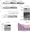
Validation of new β-TrCP substrates detected with BioID. A, Validation of β-TrCP siRNAs. Lysates from 293 T-REx cells expressing FLAGBirA*-β-TrCP1 or FLAGBirA*-β-TrCP2 in the presence of: (i) a control siRNA; (ii) two siRNAs, each directed against an individual β-TrCP paralog, or; (iii) a single siRNA directed against both β-TrCP1 and β-TrCP2 (β-TrCP1/2) were subjected to anti-FLAG Western analysis, as indicated. Actin blots are loading controls. B, Depletion of β-TrCP does not affect the half-lives of nuclear pore complex FG proteins. 293 T-REx cells were transfected with control or the indicated β-TrCP siRNAs, and treated with cycloheximide (CHX) for the indicated times. Whole cell lysates from these cells were subjected to 4–12% SDS-PAGE, and Western analysis conducted using mAb414, which recognizes FG repeat-containing nuclear pore complex proteins. C, 293 T-REx cells expressing 3xHA-NFE2L2 were transfected with control or β-TrCP siRNAs, as indicated, then treated with CHX for the indicated times. Whole cell lysates were subjected to 4–12% SDS-PAGE, then probed for NFE2L2 protein expression using an anti-HA antibody. The apparent half-life (T½) of 3xHA-NFE2L2 is indicated below each blot. D, Validation of novel β-TrCP substrate candidates. 293 T-REx cells expressing the indicated epitope-tagged proteins were transfected with control or β-TrCP siRNAs (as indicated) and treated with CHX for the indicated times. Protein expression levels were characterized using HA or FLAG antibodies. Apparent protein half-lives (in minutes) are indicated below each blot.
Fig. 3.
Fig. 3.
Stabilization of SUN2 by β-TrCP affects nuclear membrane structure. A, HeLa cells were treated with control or β-TrCP1/2 siRNA, and immunofluorescence (IF) microscopy was conducted using antibodies directed against endogenous SUN2 (red). DNA stained with DAPI (blue). B, IF of HeLa cells expressing FLAG-SUN2 and transfected with the indicated siRNA: anti-FLAG (green); anti-Lamin B (red); DAPI (blue). C, Live cell imaging of HeLa cells transiently transfected with mCherry-SUN2 and control or β-TrCP1/2 siRNA.
Fig. 4.
Fig. 4.
β-TrCP modulates P-body formation. A, HeLa Flp-In T-REx cells expressing the P-body marker GFP-DCP1A were transfected with control or β-TrCP1/2 siRNA and treated with tetracycline (1 μg/ml, 24 h) and arsenite (500 μm, 45 min), as indicated. B, Live cell confocal microscopy was conducted and P-body number quantified using Volocity software (see Experimental procedures). C, HeLa Flp-In T-REx cells expressing the stress granule marker GFP-G3BP1 were transfected with control or β-TrCP1/2 siRNA and treated with tetracycline and arsenite, as above. D, Live cell confocal microscopy was conducted and stress granule number quantified, as above. N.S.; not significant.
Fig. 5.
Fig. 5.
FLAG-PPP1R15B is stabilized by β-TrCP knockdown. A, Western blot analysis of lysates derived from 293 T-REx cells expressing FLAG-PPP1R15B treated with control or β-TrCP siRNA, as indicated. B, A PPP1R15B phosphodegron mutant is stabilized. 293 T-REx cells expressing FLAG-tagged WT or the S459–466A PPP1R15B phosphodegron mutant were treated with CHX for the indicated times. Cell lysates were subjected to Western blotting with antibodies directed against FLAG and beta actin. Apparent half lives of the FLAG-tagged PPP1R15B proteins are indicated below each blot. C, The β-TrCP interaction is disrupted in a PPP1R15B phosphodegron mutant. Co-immunoprecipitation of FLAGBirA*-β-TrCP1/2 with HA-tagged WT or S459–466A PPP1R15B proteins. D, Mutation of the phosphodegron disrupts PPP1R15B ubiquitylation. 293 T-REx cells were co-transfected with plasmids coding for FLAG-PPP1R15B WT or S459–466A and HA-ubiquitin. Lysates were prepared under denaturing conditions, diluted and FLAG IPs conducted. The isolated proteins were subjected to Western blot analysis with anti-FLAG and anti-HA. Whole cell lysates were probed with anti-HA. E, Expression of the PPP1R15B phosphodegron mutant results in decreased eIF2α Ser51 phosphorylation levels. Cell lsyates derived from 293 T-REx cells expressing FLAG-tagged WT or S459–466A PPP1R15B phosphodegron mutant proteins, and treated with vehicle (DMSO) or the ER stress agents tunicamycin or thapsigargin, were subjected to Western blotting, as indicated. F, Expression of the PPP1R15B phosphodegron mutant imparts sensitivity to ER stress agents. 293 T-REx cells expressing WT or phosphodegron mutant PPP1R15B proteins were exposed to the indicated concentrations of tunicamycin for 24 h. Viable cells, as detected by MTT assay (untreated culture numbers set to 1) are displayed (n = 3; a representative experiment is shown).

References

    1. Varshavsky A. (2012) The ubiquitin system, an immense realm. Annu. Rev. Biochem. 81, 167–176 - PubMed
    1. Hershko A., Ciechanover A. (1998) The ubiquitin system. Annu. Rev. Biochem. 67, 425–479 - PubMed
    1. Scheffner M., Staub O. (2007) HECT E3s and human disease. BMC Biochem. 8, S6. - PMC - PubMed
    1. Jiang Y. H., Beaudet A. L. (2004) Human disorders of ubiquitination and proteasomal degradation. Curr. Opin. Pediar. 16, 419–426 - PubMed
    1. Persaud A., Rotin D. (2011) Use of proteome arrays to globally identify substrates for E3 ubiquitin ligases. Methods Mol. Biol. 759, 215–224 - PubMed

Publication types

MeSH terms

LinkOut - more resources