Review
. 2015 Aug;226(2):T41-54.
doi: 10.1530/JOE-15-0113. Epub 2015 Apr 21.

60 YEARS OF NEUROENDOCRINOLOGY: The hypothalamo-pituitary-gonadal axis

Affiliations
Review

60 YEARS OF NEUROENDOCRINOLOGY: The hypothalamo-pituitary-gonadal axis

Tony M Plant. J Endocrinol. 2015 Aug.

Abstract

This review provides an outline of how our understanding of the neuroendocrine control of the hypothalamo-pituitary-gonadal axis has evolved since the publication of Geoffrey Harris' renowned monograph in 1955. Particular attention is directed to the neurobiology underlying pulsatile GnRH release from the hypothalamus, the neuroendocrine control of ovarian cycles, puberty and seasonality of gonadal function, and to ideas that have emerged as a result of examining the relationship between growth and the reproductive axis. The review closes with i) a brief discussion of how knowledge gained as a result of pursuing the early hypotheses of Harris has led to major clinical and therapeutic applications, and ii) a personal glimpse into the future of research in this fascinating area of biology.

Keywords: gonadotrophin releasing hormone; hypothalamus; neuroendocrinology; ovulation; reproduction.


Conflict of interest statement

Declaration of interest: I declare that there is no conflict of interest that could be perceived as prejudicing the impartiality of my interpretation of the research reported in this review.

Figures

Figure 1
Figure 1
Model proposed by Harris in his 1955 monograph to illustrate the relationship between the external environment and the reproductive organs (reprinted from Harris, 1955).
Figure 2
Figure 2
Demonstration of the need of intermittent GnRH stimulation of the pituitary for sustained secretion of LH (closed data points) and FSH (open data points). A monkey rendered hypogonadotropic by a hypothalamic lesion was treated initially with an intermittent iv infusion of GnRH (1 μg/min for 6 min every h). On day 0 the pulsatile regimen was terminated and replaced with a continuous GnRH infusion (1 μg/min). On day 20 the pulsatile mode of GnRH stimulation was re-instituted (reprinted from Belchetz et al, 1978).
Figure 3
Figure 3
Relationship between pulsatile secretion of GnRH (open data points) into hypophysial portal blood and corresponding episodes of pituitary LH secretion (closed data points) into the systemic circulation in an ovariectomized ewe. Modified from Clarke and Cummins, 1982 – permission pending (Copyright 1982, The Endocrine Society).
Figure 4
Figure 4
A, B. Typical morphology of mouse GnRH neurons revealed by immunohistochemistry. C. Two GnRH neurons in sheep. D. A GnRH neuron from an adult mouse has been filled in situ with the dye, biocytin, in order to facilitate visualization of the entire length of the dendrites, which in the case of the lower process is seen to extend more than 500 μm. The two higher magnification insets on the right reveal a high density of dendritic spines. Reprinted with permission from Herbison (2015).
Figure 5
Figure 5
The KNDy neuron model of the GnRH pulse generator proposed by Lehman, Coolen and Goodman. KNDy neurons reside within the arcuate nucleus (dotted purple circle) and express kisspeptin (Ks, green), neurokinin B (NKB, purple) and dynorphin (dyn, red). The model proposes that pulse generation by the network of KNDy neurons in the arcuate nucleus is achieved by a poorly understood reciprocating interplay of stimulatory NKB and inhibitory dyn inputs and an unidentified interneurone (gray). The output of the pulse generator is relayed to the GnRH neuronal network (blue) by a brief kisspeptin signal that evokes a discharge of GnRH into the hypophysial-portal circulation (shown in the lower portion of the figure). Note that the phenotype of each terminal indicates biologically relevant peptide and not selective transport of that peptide to the terminal. Similarly, the triple colored KNDy neurons indicate co-expression of the 3 peptides and not location within the cell body. RKs, Ks receptor; RNKB, NKB receptor, RDYN, dyn receptor. Reprinted with permission from Goodman and Inskeep (2015).
Figure 6
Figure 6
Chronic intermittent neurochemical stimulation of juvenile male monkeys with N-methyl DL aspartate (NMDA) readily induces a precocious pubertal pattern of pulsatile GnRH release as reflected by the emergence of corresponding discharges of LH (open data points) and testicular testosterone (closed data points) secretion. Testicular and motile epididymal sperm were typically observed after 16–26 weeks of NMDA stimulation. Means±SE (N=4) are shown. Arrows indicate time of iv injections of NMDA. Reproduced from Plant et al (1989) “Copyright (1989) National Academy of Sciences, U.S.A.”
Figure 7
Figure 7
Ovarian tissue transplanted sc to the abdomen of an adult male rhesus monkey (#7082) castrated post-pubertally and receiving anti rejection therapy with cyclosporine A exhibits regular cycles of folliculogenesis and ovulation followed by a normal luteal phase. Numbers on the peaks in LH indicate maximum concentration of the gonadotropin on that day. Modified from Norman and Spies (1986) with permission (Copyright 1986, The Endocrine Society).
Figure 8
Figure 8
A model for the seasonal control of pulsatile GnRH release. The duration of the photoperiod is relayed by melatonin to melatonin receptors (MT1) in the thyrotrophs of the pars tuberalis (PT), and further relayed by thyrotrophin (TSH) to the mediobasal hypothalamus, where the arcuate nucleus KNDy neurons are located. TSH upregulates the expression of the genes encoding deiodinase 2 and 3 (DIO2 and DIO3), in specialized ependymal cells (tanycytes) lining the base of the third ventricle (3v). The diodinase enzymes convert thyroid hormone (T4) into the active metabolite, tri-iodothyronine (T3), and the increase in thyroid hormone activity dictates the level of GnRH pulsatility, which in turn governs the gonadotrophin output from the gonadotrophs in the pars distalis (PD). TH is considered to reach the MBH via the 3v and/or from brain capillaries (Cp). Reprinted with permission from Hazelrigg and Simonneaux (2015).

References

    1. Amoss M, Burgus R, Blackwell R, Vale W, Fellows R, Guillemin R. Purification, amino acid composition and N-terminus of the hypothalamic luteinizing hormone releasing factor (LRF) of ovine origin. Biochem Biophys Res Commun. 1971;44:205–210. - PubMed
    1. Barry J, Dubois MP, Poulain P. LRF producing cells of the mammalian hypothalamus. A fluorescent antibody study. Z Zellforsch Mikrosk Anat. 1973;146:351–366. - PubMed
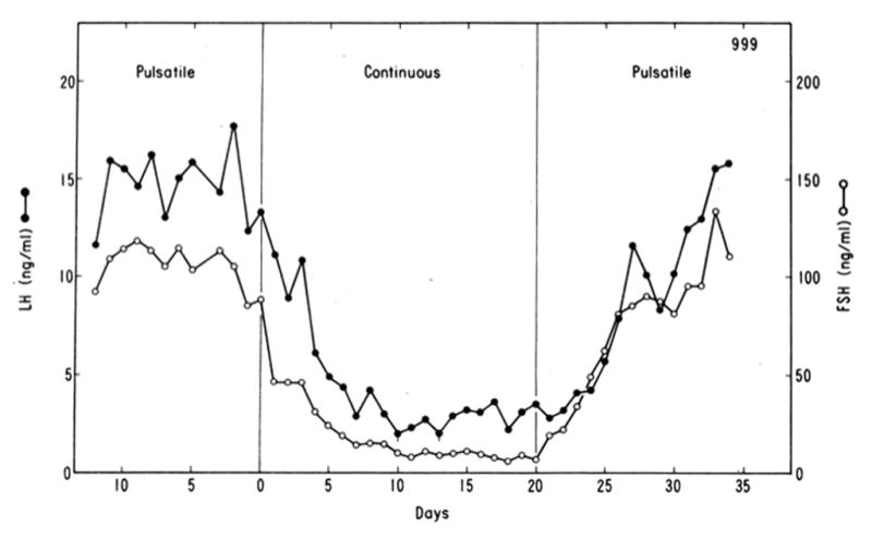
    1. Belchetz PE, Plant TM, Nakai Y, Keogh EJ, Knobil E. Hypophysial responses to continuous and intermittent delivery of hypopthalamic gonadotropin-releasing hormone. Science. 1978;202:631–633. - PubMed
    1. Cheng G, Coolen LM, Padmanabhan V, Goodman RL, Lehman MN. The kisspeptin/neurokinin B/dynorphin (KNDy) cell population of the arcuate nucleus: sex differences and effects of prenatal testosterone in sheep. Endocrinology. 2010;151:301–311. - PMC - PubMed
    1. Clark SJ, Ellis N, Styne DM, Gluckman PD, Kaplan SL, Grumbach MM. Hormone ontogeny in the ovine fetus. XVII. Demonstration of pulsatile luteinizing hormone secretion by the fetal pituitary gland. Endocrinology. 1984;115:1774–1779. - PubMed

Publication types