

Primary cilia modulate balance of canonical and non-canonical Wnt signaling responses in the injured kidney
- PMID: 25901180
- PMCID: PMC4404279
- DOI: 10.1186/s13069-015-0024-y
Primary cilia modulate balance of canonical and non-canonical Wnt signaling responses in the injured kidney
Abstract
Background: While kidney injury is associated with re-expression of numerous Wnt ligands and receptors, molecular mechanisms which underlie regulation of distinct Wnt signaling pathways and ensuing biological consequences remain incompletely understood. Primary cilia are increasingly being recognized as cellular 'antennae' which sense and transduce signals from the microenvironment, particularly through Wnt signaling. Here, we explored the role of cilia as modulators of canonical and non-canonical Wnt signaling activities involving tubular epithelial cells in the injured kidney.
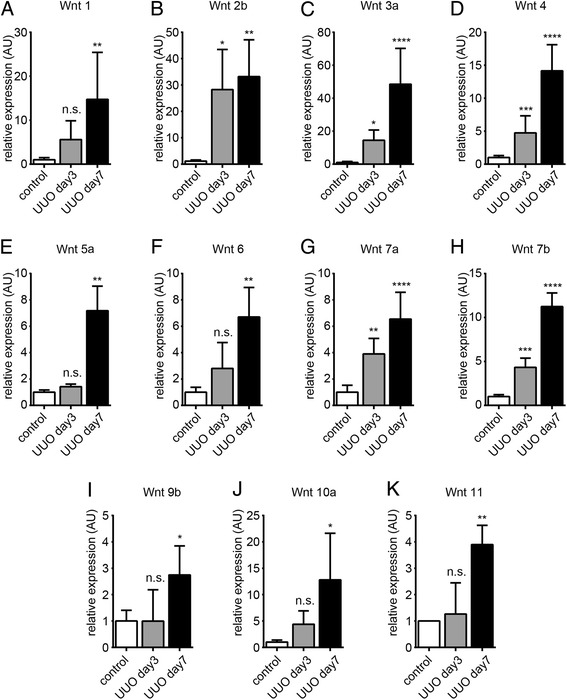
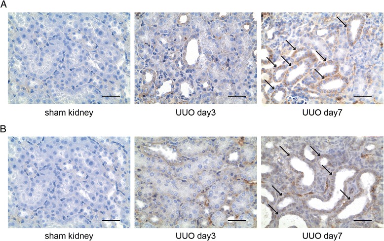
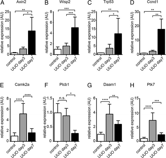
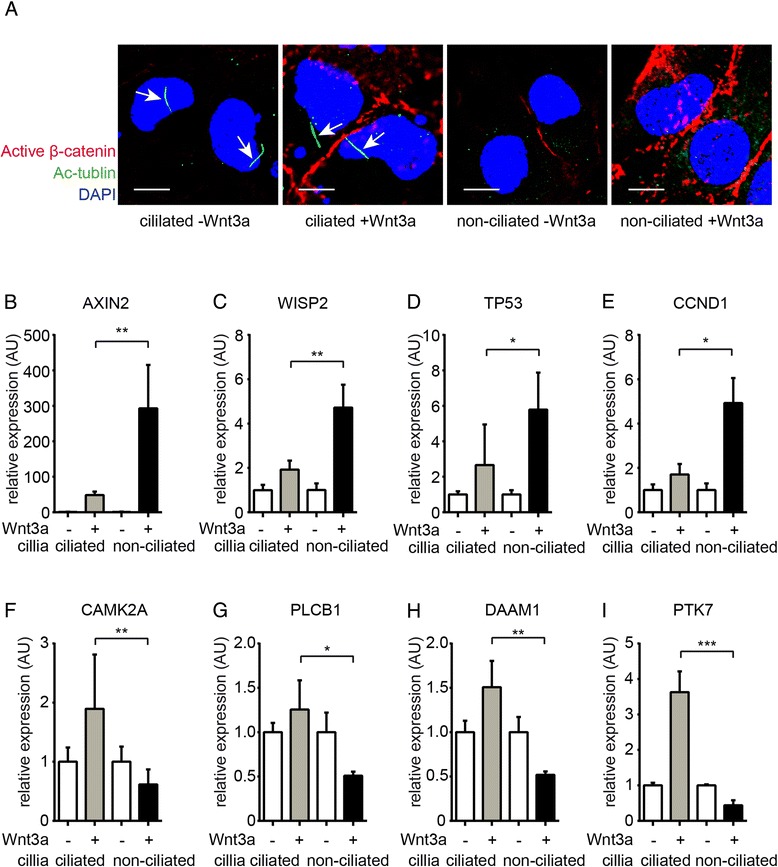
Results: We demonstrate that in the mouse model of unilateral ureter obstruction, progression of kidney injury correlates with increased expression of numerous Wnt ligands, and that increased expression of Wnt ligands corresponded with over-activation of canonical Wnt signaling. In contrast, non-canonical Wnt signaling dropped significantly during the course of kidney injury despite gradually increased expression of typical non-canonical and intermediate Wnt signaling ligands. We further demonstrate that in cultured tubular epithelial cells, cilia modulate balance between canonical and non-canonical signaling responses upon exposure to Wnt ligands.
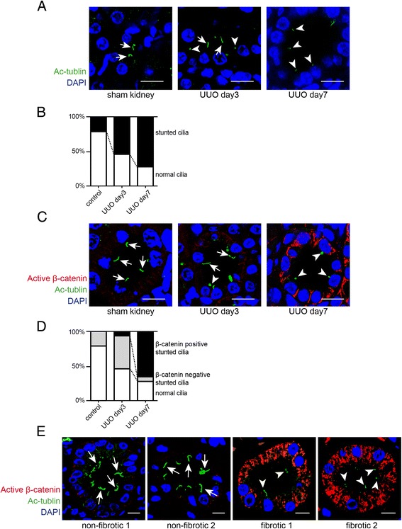
Conclusions: We provide evidence that in the context of renal injury, primary cilia act as molecular switches between canonical and non-canonical Wnt signaling activity, possibly determining between regenerative and pro-fibrotic effects of Wnt re-expression in the injured kidney.
Keywords: Cilia; Epithelial; Fibrosis.
Figures
Similar articles
-
Wnt-dependent beta-catenin signaling is activated after unilateral ureteral obstruction, and recombinant secreted frizzled-related protein 4 alters the progression of renal fibrosis.J Am Soc Nephrol. 2005 Aug;16(8):2373-84. doi: 10.1681/ASN.2004110949. Epub 2005 Jun 8. J Am Soc Nephrol. 2005. PMID: 15944336
-
Wnt and planar cell polarity signaling in cystic renal disease.Organogenesis. 2014 Jan 1;10(1):86-95. doi: 10.4161/org.26766. Epub 2013 Oct 25. Organogenesis. 2014. PMID: 24162855 Free PMC article. Review.
-
Wnt-4 activates the canonical beta-catenin-mediated Wnt pathway and binds Frizzled-6 CRD: functional implications of Wnt/beta-catenin activity in kidney epithelial cells.Exp Cell Res. 2004 Aug 15;298(2):369-87. doi: 10.1016/j.yexcr.2004.04.036. Exp Cell Res. 2004. PMID: 15265686
-
Connective tissue growth factor induces tubular epithelial to mesenchymal transition through the activation of canonical Wnt signaling in vitro.Ren Fail. 2015 Feb;37(1):129-35. doi: 10.3109/0886022X.2014.967699. Epub 2014 Oct 8. Ren Fail. 2015. PMID: 25296105
-
Novel biomarkers in kidney disease: roles for cilia, Wnt signalling and ATMIN in polycystic kidney disease.Biochem Soc Trans. 2016 Dec 15;44(6):1745-1751. doi: 10.1042/BST20160124. Biochem Soc Trans. 2016. PMID: 27913685 Review.
Cited by
-
Expression Pattern of α-Tubulin, Inversin and Its Target Dishevelled-1 and Morphology of Primary Cilia in Normal Human Kidney Development and Diseases.Int J Mol Sci. 2021 Mar 28;22(7):3500. doi: 10.3390/ijms22073500. Int J Mol Sci. 2021. PMID: 33800671 Free PMC article.
-
Progression of chronic kidney disease: too much cellular talk causes damage.Kidney Int. 2017 Mar;91(3):552-560. doi: 10.1016/j.kint.2016.08.025. Epub 2016 Oct 20. Kidney Int. 2017. PMID: 27773427 Free PMC article. Review.
-
Disturbances in Switching between Canonical and Non-Canonical Wnt Signaling Characterize Developing and Postnatal Kidneys of Dab1-/- (yotari) Mice.Biomedicines. 2023 Apr 28;11(5):1321. doi: 10.3390/biomedicines11051321. Biomedicines. 2023. PMID: 37238991 Free PMC article.
-
Can Tissue Cilia Lengths and Urine Cilia Proteins Be Markers of Kidney Diseases?Chonnam Med J. 2018 May;54(2):83-89. doi: 10.4068/cmj.2018.54.2.83. Epub 2018 May 25. Chonnam Med J. 2018. PMID: 29854673 Free PMC article. Review.
-
Identification of New Ciliary Signaling Pathways in the Brain and Insights into Neurological Disorders.J Neurosci. 2025 Aug 13;45(33):e0800242025. doi: 10.1523/JNEUROSCI.0800-24.2025. J Neurosci. 2025. PMID: 40623838 Free PMC article.
References
LinkOut - more resources
Full Text Sources
Other Literature Sources
