Review
. 2015 Apr 16;7(4):318-27.
doi: 10.4253/wjge.v7.i4.318.

Endoscopic ultrasound in the evaluation of pancreatic neoplasms-solid and cystic: A review

Affiliations
Review

Endoscopic ultrasound in the evaluation of pancreatic neoplasms-solid and cystic: A review

Eric M Nelsen et al. World J Gastrointest Endosc. .

Abstract

Pancreatic neoplasms have a wide range of pathology, from pancreatic adenocarcinoma to cystic mucinous neoplasms. Endoscopic ultrasound (EUS) with or without fine needle aspiration (FNA) is a helpful diagnostic tool in the work-up of pancreatic neoplasms. Its utility in pancreatic malignancy is well known. Over the last two decades EUS-FNA has become a procedure of choice for diagnosis of pancreatic adenocarcinoma. EUS-FNA is highly sensitive and specific for solid lesions, with sensitivities as high as 80%-95% for pancreatic masses and specificity as high as 75%-100%. Multiple aspects of the procedure have been studied to optimize the rate of diagnosis with EUS-FNA including cytopathologist involvement, needle size, suctioning and experience of endoscopist. Onsite pathology is one of the most important elements in increasing diagnostic yield rate in EUS-FNA. EUS-FNA is valuable in diagnosing rare and atypical pancreatic neoplasms including neuroendocrine, lymphoma and metastatic disease. As more and more patients undergo cross sectional imaging, cystic lesions of the pancreas are becoming a more common occurrence and EUS-FNA of these lesions can be helpful for differentiation. This review covers the technical aspects of optimizing pancreatic neoplasm diagnosis rate, highlight rare pancreatic neoplasms and role of EUS-FNA, and also outline the important factors in diagnosis of cystic lesions by EUS-FNA.

Keywords: Endoscopic ultrasound-fine needle aspiration; Pancreatic adenocarcinoma; Pancreatic cysts; Pancreatic neoplasms; Review.


Figures

Figure 1
Figure 1
Pancreatic adenocarcinoma. A: Endoscopic ultrasound image demonstrating a large pancreatic adenocarcinoma; B: Pancreatic adenocarcinoma. A crowded group of large, pleomorphic ductal cells with irregular hyperchromatic nuclei and prominent anisocytosis. These contrast well with an orderly sheet of benign ductal epithelial cells with round, uniform nuclei (bottom) (Diff-QuikTM stain, × 100); C: Similar in appearance malignant cells in a Papanicolaou-stained preparation (× 400).
Figure 2
Figure 2
Pancreatic neuroendocrine neoplasm. A: Endoscopic ultrasound image showing a 9 mm × 10 mm neuroendocrine tumor (insulinoma); B: Low-power view shows a cellular aspirate composed of clusters of uniform cells (Diff-QuikTM stain, × 100); C: High power view shows uniform cells with high N:C ratios and coarse chromatin (Diff-QuikTM stain, × 400); D: Papanicolaou stain highlights coarse, evenly distributed chromatin (× 400).
Figure 3
Figure 3
Primary pancreatic lymphoma. A: Endoscopic ultrasound demonstrating a 1.8 cm × 2.2 cm lymphoma in the uncinate process of the pancreas; B: Low-power view showing a very cellular aspirate composed of discohesive lymphoid cells (Diff-QuikTM stain, × 100); C: High-power view showing an admixture of mature lymphocytes of various sizes with no more than a minimal atypia; lymphoid aggregates resembling a germinal center are also present (bottom); D: Small mature lymphocytes with cleaved and irregular nuclei raising suspicion for a mature B-cell lymhoma. (Diff-QuikTM stain, × 400).
Figure 4
Figure 4
Endoscopic ultrasound image of large, 3.5 cm × 4.4 cm, round, hypoechoic, heterogenous mass lesion arising from the tail of the pancreas.
Figure 5
Figure 5
Cytology from a primary pancreatic gastrointestinal stromal tumor.
Figure 6
Figure 6
Pancreatic gastrointestinal stromal tumor, cytology demonstrates a spindle cell neoplasm with moderate nuclear pleomorphism which stains strongly positive for CD117 and negative for desmin, consistent with a gastrointestinal stromal tumor arising from the pancreas. (Courtesy of Rashmi Agni, University of Wisconsin Department of Pathology and Laboratory Medicine).
Figure 7
Figure 7
Metastatic high-grade serous carcinoma of the ovary. A: Endoscopic ultrasound image of a metastatic high-grade serous carcinoma of the ovary; B: Low-power view showing a cellular aspirate with a necrotic background (Diff-QuikTM stain, × 100); C: High-power view showing groups of malignant cells with large nuclei and prominent nucleoli. These cells are difficult to distinguish from a primary pancreatic ductal adenocarcinoma; however, necrotic background is not common in a primary tumor (Diff-QuikTM stain, × 400); D: Papanicolaou stain showing a cannon ball shaped group of malignant cells with large, round nuclei and prominent nucleoli, characteristic of serous ovarian carcinoma (× 400).
Figure 8
Figure 8
Endoscopic ultrasound image demonstrating a cystadenocarcinoma.
Figure 9
Figure 9
Endoscopic ultrasound image demonstrating an intraductal papillary-mucinous neoplasm.
Figure 10
Figure 10
Endoscopic view of “fish mouth papilla” due to the presence of mucin within the main duct.
Figure 11
Figure 11
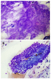
Pancreatic mucinous neoplasm. A: Low-power view of pancreatic mucinous neoplasm showing copious thick, colloid-like mucin (Diff-QuikTM stain, × 100); B: High-power view of pancreatic mucinous neoplasm showing sheets of only mildly atypical columnar cells containing intracytoplasmic mucin; these cells are very difficult to distinguish from benign gastric or duodenal epithelium (Diff-QuikTM stain, × 400).

Similar articles

Cited by

References

    1. Helmstaedter L, Riemann JF. Pancreatic cancer--EUS and early diagnosis. Langenbecks Arch Surg. 2008;393:923–927. - PubMed
    1. Jemal A, Siegel R, Ward E, Hao Y, Xu J, Thun MJ. Cancer statistics, 2009. CA Cancer J Clin. 2009;59:225–249. - PubMed
    1. Volmar KE, Vollmer RT, Jowell PS, Nelson RC, Xie HB. Pancreatic FNA in 1000 cases: a comparison of imaging modalities. Gastrointest Endosc. 2005;61:854–861. - PubMed
    1. Dewitt J, Devereaux BM, Lehman GA, Sherman S, Imperiale TF. Comparison of endoscopic ultrasound and computed tomography for the preoperative evaluation of pancreatic cancer: a systematic review. Clin Gastroenterol Hepatol. 2006;4:717–725; quiz 664. - PubMed
    1. Vilmann P, Jacobsen GK, Henriksen FW, Hancke S. Endoscopic ultrasonography with guided fine needle aspiration biopsy in pancreatic disease. Gastrointest Endosc. 1992;38:172–173. - PubMed