

Cervical paravertebral osteolipoma: case report and literature review
- PMID: 25901244
- PMCID: PMC4404547
- DOI: 10.4184/asj.2015.9.2.290
Cervical paravertebral osteolipoma: case report and literature review
Abstract
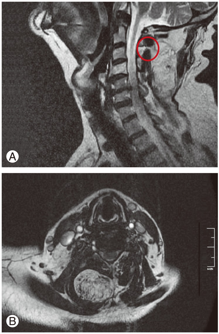
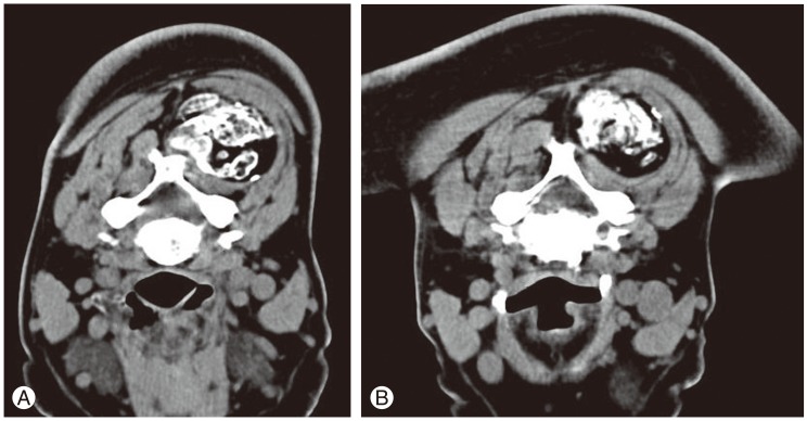
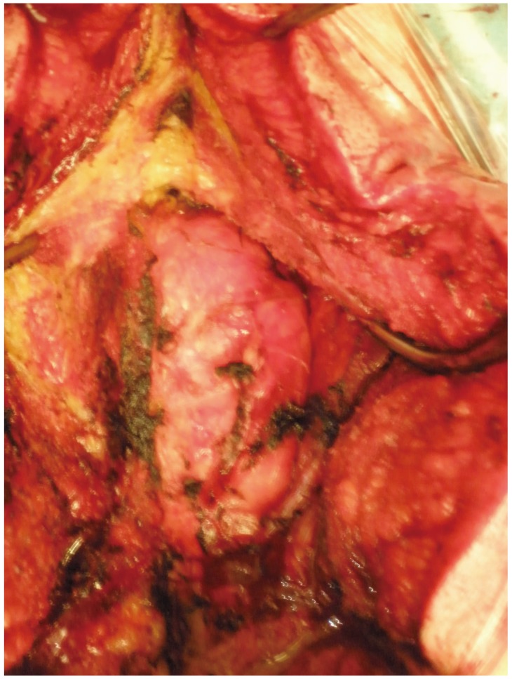
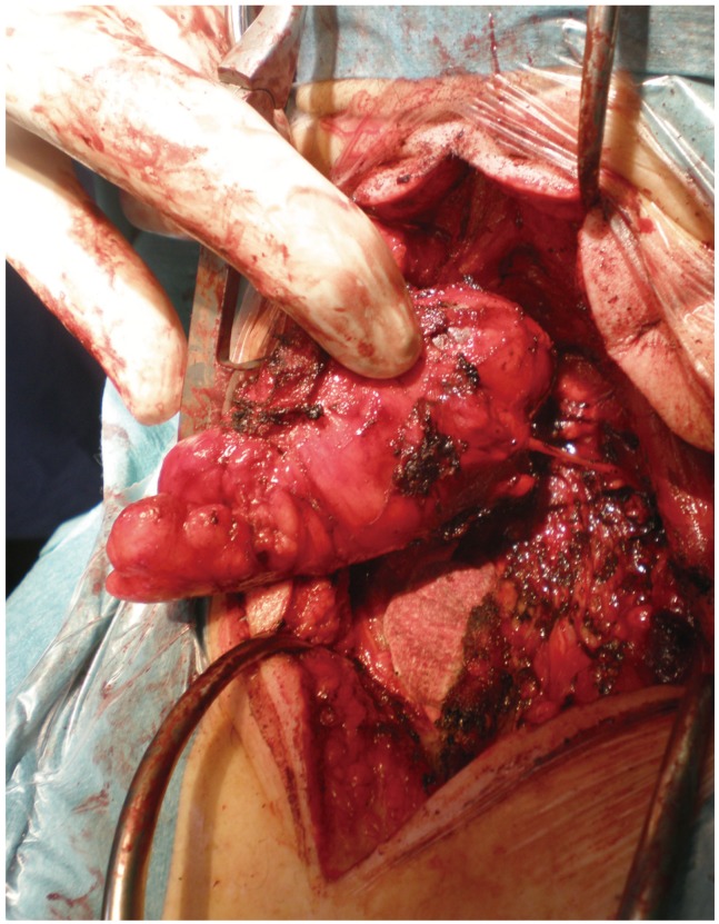
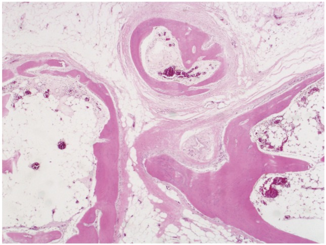
Lipomas are the most frequent soft tissue tumors. Osteolipomas are a rare variant that can be difficult to diagnose. We report the case of a 66-year-old man consulting with a tumor of 2 years development in the right paravertebral cervical region. Neurologically, the patient had no sign of myelopathy or neurological focality. Magnetic resonance imaging showed a mass with a lipid component and calcifications inside within the right paravertebral musculature with a possible origin in the right C3 posterior root. A computed tomography scan and guided biopsy were performed, revealing hematic material and small bone spicules with no apparent neoplastic element. The tumor was totally removed, including the right C3 posterior branch, and was confirmed to be an osteolipoma on biopsy. The patient remains asymptomatic at 6-month follow-up. The osteolipoma is a benign tumor of soft tissue, characterized by lipoma areas with mature bone tissue differentiation, and even with hematopoietic marrow.
Keywords: Atypical lipoma; Cervical paravertebral; Ossifying lipoma; Soft tissue tumor.
Conflict of interest statement
Figures
References
-
- Allen PW. Tumors and proliferations of adipose tissue: a clinicopathologic approach. New York: Masson Pub., USA; 1981.
-
- Obermann EC, Bele S, Brawanski A, Knuechel R, Hofstaedter F. Ossifying lipoma. Virchows Arch. 1999;434:181–183. - PubMed
-
- Kameyama K, Akasaka Y, Miyazaki H, Hata J. Ossifying lipoma independent of bone tissue. ORL J Otorhinolaryngol Relat Spec. 2000;62:170–172. - PubMed
-
- Bohm KC, Birman MV, Silva SR, et al. Ossifying lipoma of c1-c2 in an adolescent. J Pediatr Orthop. 2011;31:e53–e56. - PubMed
-
- Bulkeley W, Mills OL, Gonzalvo A, Wong K. Osteolipoma of the parapharyngeal space mimicking liposarcoma: a case report. Head Neck. 2012;34:301–303. - PubMed
LinkOut - more resources
Full Text Sources
Other Literature Sources
Miscellaneous
