. 2015 Apr 22;10(4):e0124395.
doi: 10.1371/journal.pone.0124395. eCollection 2015.

Targets of Wnt/ß-catenin transcription in penile carcinoma

Affiliations

Targets of Wnt/ß-catenin transcription in penile carcinoma

Manit Arya et al. PLoS One. .

Abstract

Penile squamous cell carcinoma (PeCa) is a rare malignancy and little is known regarding the molecular mechanisms involved in carcinogenesis of PeCa. The Wnt signaling pathway, with the transcription activator ß-catenin as a major transducer, is a key cellular pathway during development and in disease, particularly cancer. We have used PeCa tissue arrays and multi-fluorophore labelled, quantitative, immunohistochemistry to interrogate the expression of WNT4, a Wnt ligand, and three targets of Wnt-ß-catenin transcription activation, namely, MMP7, cyclinD1 (CD1) and c-MYC in 141 penile tissue cores from 101 unique samples. The expression of all Wnt signaling proteins tested was increased by 1.6 to 3 fold in PeCa samples compared to control tissue (normal or cancer adjacent) samples (p<0.01). Expression of all proteins, except CD1, showed a significant decrease in grade II compared to grade I tumors. High magnification, deconvolved confocal images were used to measure differences in co-localization between the four proteins. Significant (p<0.04-0.0001) differences were observed for various permutations of the combinations of proteins and state of the tissue (control, tumor grades I and II). Wnt signaling may play an important role in PeCa and proteins of the Wnt signaling network could be useful targets for diagnosis and prognostic stratification of disease.


Conflict of interest statement

Competing Interests: AA (principal inventor) and CT and JRM (co-inventors) have filed for a patent through UCL Business for targeting Wnt signaling pathway for ß-catenin related disorders (New British Patent Application no. 1318659.8). AA is an associate editor of PLOS ONE. These do not alter the authors' adherence to all the PLOS ONE policies and to sharing data and materials.

Figures

Fig 1
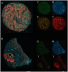
Fig 1. Representative images from Zeiss AxioScan Z1 slide scanner.
Composite, overlay, of four fluorophores (FITC, Alexa Fluor-405, Cy3 and Cy5 for MMP7, Wnt4, CD1 and c-MYC, respectively) in (A) PeCa tissue cores (core B2 of PE241) and (B) control (core D5 of PE241, see methods). C-F are individual images used to construct the composite of PeCa tissue core, for MMP7 (C, green), Wnt4 (D, blue), CD1 (E, orange) and c-MYC (F, red). G-J are individual images used to construct the composite of control tissue core, for MMP7 (G, green), Wnt4 (H, blue), CD1 (I, orange) and c-MYC (J, red). Quantitative analysis was performed on the non-enhanced, original, gray images for which all settings were identical in all tissue core images analyzed. Scale bar = 250 μm.
Fig 2
Fig 2. Box plot of the quantitation of expression of WNT4, MMP7, CD1 and c-MYC in PeCa tissue classified according to pathological grade.
97% of the samples used in this study were classified as grade I or II. The data presented in Table 1 was segregated into expression according to pathological grades and plotted as box plots. Filled circles are 99% value and horizontal line within the box is the median value. Protein expression (mean gray value per core) in control samples (green, n = 37); malignant tumor sample classified as grade I by a pathologist (blue, n = 82) or grade II (purple, n = 18). All proteins showed a significant difference in expression between control and malignant tumor samples. Significance of difference was measured using Mann-Whitney U test: * = p<0.001 and § = p<0.01 between control and malignant grade I or grade II samples or within grade I and grade II. There was no significant difference in protein expression for any marker between grade I and grade II samples.
Fig 3
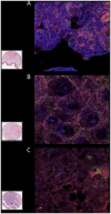
Fig 3. High magnification deconvolved images for WNT4, MMP7, CD1 and c-MYC co-expression in control and malignant penile tissue.
Composite, representative, micrographs of immunofluorescently stained penile tissue cores used to quantify co-localization of four proteins WNT4 (Alexa 405-blue), MMP7 (FITC-green), CD1 (Cy3-orange) and c-MYC (Cy5-red) after deconvolution. Tissue cores from 8 patients (control), 25 malignant tissue cores (11 grade I and 14 grade II) were imaged using an Olympus FV1000 confocal microscope. High magnification imaging (at 1024x1024 resolution) was performed for deconvolution analysis using Huygens Profesional software. Areas of squamous cell carcinoma in malignant tissue cores were identified, visually (see box) from a serial H&E section of the tissue, and imaged at high magnification (40x with 4–6x zoom). Two representative immunofluorescently labelled cores are shown: A is control and B and C are grade I and grade II, squamous cell carcinoma samples, respectively; Inset is H&E serial sections of at low magnification. High magnification images were deconvolved to reduce noise and converted to 16bit tif files for each fluorophore (Alexa Fluor-405, FITC, Cy3 and Cy5) were imported into Huygens deconvolution software and pseudo colored (FITC, green; Alexa Fluor-405, blue; Cy3, orange; and Cy5, red). Z-projections for up to 53 images for each tissue core. Representative images of controls and malignant carcinoma samples are shown. Scale bar = 3μm. High magnification images were used for quantitative co-localization measurements.
Fig 4
Fig 4. Box plot of differential pattern for WNT4, MMP7, CD1 and c-MYC co-expression in control and malignant penile tissue.
Quantitative co-localization was performed from high magnification images (e.g. as represented in Fig 3) and image files used for quantitation as described in materials and methods in WCIF ImageJ software co-localization plugin to measure the Pearson coefficient of co-localization. 35 individual tissue cores that included control (gradient from centre formula image) and malignant grade I (gradient from left to right formula image) and grade II (gradient from right to left formula image) were used for each fluorophore to calculate the co-localization of expression of WNT4, MMP7, CD1 and c-MYC, generating 12 comparisons: CD1 and WNT4 (orange/blue), MMP7 and WNT4 (green/blue), c-MYC and WNT4 (pink/blue), CD1 and c-MYC (orange/pink), MMP7 and c-MYC (green/pink), MMP7 and CD1 (green/orange). Significance of difference in the Pearson coefficient of co-localization between control and grades I and II (*0.02–0.04) and between grade I and grade II (§ p<0.04, range 0.04–0.0001) samples was calculated using the Mann-Whitney U test; boxes showing comparisons without symbols are not significantly different.

Similar articles

Cited by

References

    1. Pizzocaro G, Algaba F, Horenblas S, Solsona E, Tana S, et al. (2010) EAU penile cancer guidelines 2009. Eur Urol 57: 1002–1012. S0302-2838(10)00105-3 [pii]; 10.1016/j.eururo.2010.01.039 - DOI - PubMed
    1. Arya M, Kalsi J, Kelly J, Muneer A (2013) Malignant and premalignant lesions of the penis. BMJ 346: f1149 10.1136/bmj.f1149 - DOI - PubMed
    1. Arya M, Li R, Pegler K, Sangar V, Kelly JD, et al. (2013) Long-term trends in incidence, survival and mortality of primary penile cancer in England. Cancer Causes Control 24: 2169–2176. 10.1007/s10552-013-0293-y - DOI - PubMed
    1. Martins AC, Faria SM, Cologna AJ, Suaid HJ, Tucci S Jr (2002) Immunoexpression of p53 protein and proliferating cell nuclear antigen in penile carcinoma. J Urol 167: 89–92. S0022-5347(05)65389-X [pii]. - PubMed
    1. Mentrikoski MJ, Stelow EB, Culp S, Frierson HF Jr, Cathro HP (2014) Histologic and immunohistochemical assessment of penile carcinomas in a North American population. Am J Surg Pathol 38: 1340–1348. 10.1097/PAS.0000000000000124;00000478-201410000-00005 [pii]. - DOI - PubMed

Publication types

LinkOut - more resources