. 2015 Apr 22:6:6945.
doi: 10.1038/ncomms7945.

ASK1 restores the antiviral activity of APOBEC3G by disrupting HIV-1 Vif-mediated counteraction

Affiliations

ASK1 restores the antiviral activity of APOBEC3G by disrupting HIV-1 Vif-mediated counteraction

Kei Miyakawa et al. Nat Commun. .

Abstract

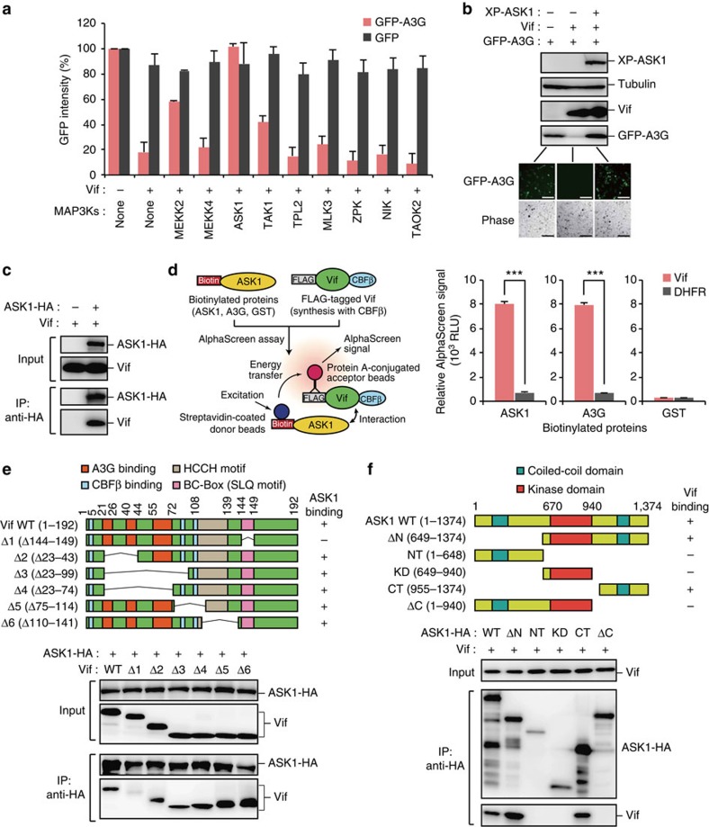
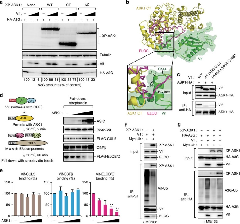
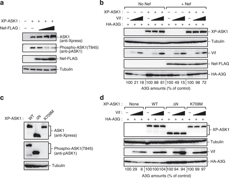
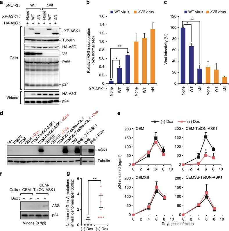
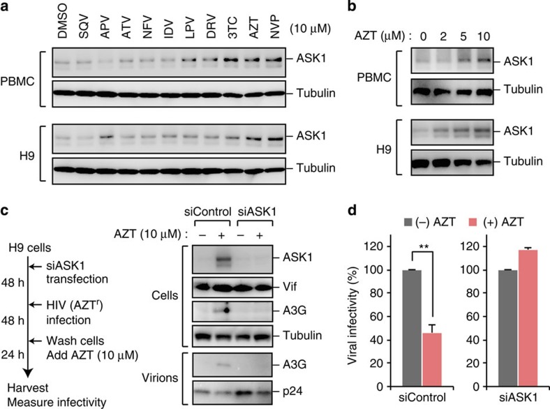
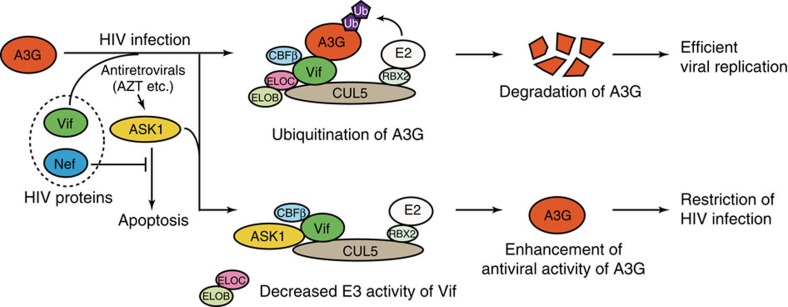
APOBEC3G (A3G) is an innate antiviral restriction factor that strongly inhibits the replication of human immunodeficiency virus type 1 (HIV-1). An HIV-1 accessory protein, Vif, hijacks the host ubiquitin-proteasome system to execute A3G degradation. Identification of the host pathways that obstruct the action of Vif could provide a new strategy for blocking viral replication. We demonstrate here that the host protein ASK1 (apoptosis signal-regulating kinase 1) interferes with the counteraction by Vif and revitalizes A3G-mediated viral restriction. ASK1 binds the BC-box of Vif, thereby disrupting the assembly of the Vif-ubiquitin ligase complex. Consequently, ASK1 stabilizes A3G and promotes its incorporation into viral particles, ultimately reducing viral infectivity. Furthermore, treatment with the antiretroviral drug AZT (zidovudine) induces ASK1 expression and restores the antiviral activity of A3G in HIV-1-infected cells. This study thus demonstrates a distinct function of ASK1 in restoring the host antiviral system that can be enhanced by AZT treatment.


Figures

Figure 1
Figure 1. ASK1 binds and counteracts Vif.
(a,b) ASK1 inhibits Vif-mediated A3G degradation. HEK293 cells were cotransfected with plasmids encoding Vif (250 ng), GFP-A3G (50 ng) and the indicated HA-tagged MAP3Ks or Xpress (XP)-tagged ASK1 (500 ng). After 48 h, GFP intensities (n=3, mean±s.d.), cell images and protein expression levels were analysed by flow cytometry (a), fluorescence microscopy and western blotting (b), respectively. Scale bar, 100 μm. (c) ASK1 interacts with Vif in cells. Cells were cotransfected with plasmids encoding ASK1-HA and Vif. Cell lysates were immunoprecipitated with an anti-HA antibody and the bound proteins were then analysed by western blotting. (d) In vitro interaction of ASK1 with Vif. Schematic representation of the amplified luminescent proximity homogeneous (AlphaScreen) assay used to detect the direct protein–protein interaction (left). In this study, FLAG-tagged Vif proteins were co-expressed with CBFβ to stabilize the conformation of the Vif protein. AlphaScreen analysis (n=3, mean±s.d.) using recombinant ASK1, A3G and Vif proteins is shown (right). GST and DHFR (dihydrofolate reductase) were used as negative controls. ***P<0.001, two-tailed unpaired t-test. (e) HEK293 cells were cotransfected with plasmids encoding ASK1-HA and Vif or one of its truncated mutants (top). Cell lysates were immunoprecipitated with anti-HA antibody and bound Vif proteins were detected with western blot analysis (bottom). (f) HEK293 cells were cotransfected with plasmids encoding Vif and ASK1-HA or one of its truncated mutants (top). Cell lysates were immunoprecipitated with anti-HA antibody and bound Vif proteins were detected by western blot analysis (bottom). Full images for all western blots analysis are shown in Supplementary Fig. 5.
Figure 2
Figure 2. ASK1 inhibits the formation of Vif–E3 ubiquitin ligase complex.
(a) The C-terminal domain (CT) of ASK1 is sufficient to impair Vif-mediated A3G degradation. HEK293 cells were cotransfected with plasmids encoding XP-ASK1 (500 ng), HA-A3G (10 ng) and Vif (50 or 100 ng). Protein expression was then detected using western blot analysis. The numerical values below the blot indicate the amounts of HA-A3G determined with densitometry. (b) The structural model of ASK1 CT (yellow) was generated and subjected to docking simulation with Vif (green, PDB: 4N9F). ELOC (purple) was overlaid onto the Vif–ASK1 model. The square shows the predicted inhibition by ASK1 of ELOC binding to the BC-Box of Vif. (c) The BC-box motif of Vif is important for the binding of ASK1. HEK293 cells were cotransfected with plasmids encoding ASK1-HA and Vif or the indicated mutants. Cell lysates were immunoprecipitated with anti-HA antibody and bound Vif proteins were detected by western blot analysis. (d,e) ASK1 inhibits the formation of the ubiquitin ligase complex by blocking Vif interaction with ELOB/C. (d) Recombinant biotinylated Vif was co-synthesized with CBFβ and then pre-mixed with various amounts of ASK1 for 5 min before the addition of equivalent amounts of FLAG-tagged CUL5, ELOB and ELOC. After 1 h, the mixture was processed for pull down with streptavidin-coated magnetic beads as shown on the left. Bound proteins were detected by western blot (right panels). The bar charts shown in e indicate the amounts of Vif-associated proteins in the presence of ASK1, determined by densitometric analysis of the western blots (n=3, mean±s.d.). **P<0.01, two-tailed unpaired t-test. (f) HEK293 cells were cotransfected with plasmids encoding XP-ASK1, Vif and Myc-Ubiquitin (Ub). Cells were then treated with MG132 for 18 h before being harvested. Cell lysates were immunoprecipitated with anti-Vif antibody and the ligase activity of Vif was detected by western blot analysis using an anti-Myc antibody. (g) HEK293 cells were cotransfected with plasmids encoding XP-ASK1, HA-A3G, Vif and Myc-Ub. Cells were then treated with MG132 for 18 h before being harvested. Cell lysates were immunoprecipitated with anti-HA antibody and the Vif-induced ubiquitination of A3G was detected by western blot analysis using anti-Myc antibody. Full images for all western blots analysis are shown in Supplementary Fig. 5.
Figure 3
Figure 3. Nef does not affect ASK1-mediated Vif inactivation.
(a) Nef inhibits the autophosphorylation of ASK1. HEK293 cells were cotransfected with plasmids encoding XP-ASK1 (100 ng) and Nef-FLAG (100 or 200 ng). Cell lysates were subjected to western blot analysis using the indicated antibodies. (b) Nef does not alter the effect of ASK1 on Vif-mediated A3G degradation. HEK293 cells were cotransfected with plasmids encoding XP-ASK1 (500 ng), HA-A3G (10 ng), Vif (50 or 100 ng) and Nef-FLAG (1 μg). Protein expression was detected by western blot. The numerical values below the blot indicate the amounts of HA-A3G determined with densitometry. (c) HEK293 cells were transfected with the indicated ASK1 mutants (ΔN, constitutively active; K709M, kinase-dead). Cell lysates were subjected to western blotting against the indicated antibodies. (d) HEK293 cells were cotransfected with plasmids encoding HA-A3G (10 ng), Vif (50 or 100 ng) and with wild-type ASK1 or one of its kinase mutants (500 ng). Protein expression was detected by western blot. The numerical values below the blot indicate the amounts of HA-A3G as determined by densitometry. Full images for all western blots analysis are shown in Supplementary Fig. 5.
Figure 4
Figure 4. ASK1 restricts HIV-1 replication via A3G reactivation.
(ac) ASK1 expression in virus-producing cells promotes A3G incorporation into virions and reduces infectivity. HEK293 cells were cotransfected with an HIV-1 molecular clone carrying a GFP reporter gene (pNL4-3ΔEnv-GFP) or its Vif-deficient mutant (pNL4-3ΔEnvΔVif-GFP) together with a plasmid encoding VSV-G, XP-ASK1 and HA-A3G. (a,b) Forty-eight hours after transfection, cell lysates and supernatants were harvested and analysed by western blotting against the indicated antibodies. The bar chart in b indicates the amounts of HA-A3G normalized by p24 levels in virions, as determined by densitometric analysis of western blots (n=3, mean±s.d.). (c) The CD4+ T-cell line (M8166) was infected with harvested and normalized virus for 2 days and infected (GFP-positive) cells were then measured by flow cytometry (n=3, mean±s.d.). *P<0.05; **P<0.01, two-tailed unpaired t-test. (d) Expression levels of ASK1 in DOX-treated or untreated CEM-TetON-ASK1 and CEMSS-TetON-ASK1 cells. 293 cells transfected with ASK1 or treated with PMA are shown as positive controls. (eg) CEM-TetON-ASK1 and CEMSS-TetON-ASK1 cells and their parent cell lines (CEM and CEMSS) were infected with HIV-1. (e) Culture supernatants were harvested at the indicated time-points and subjected to p24 ELISA (n=3, mean±s.d.). *P<0.05, two-tailed unpaired t-test. (f) The incorporation of A3G into virions from indicated cells (at 8 d.p.i.) was detected by western blot. (g) The infected cells (CEM-TetON-ASK1) were harvested at 8 d.p.i. and subjected to G-to-A hypermutation analysis (n=8, mean±s.d.). **P<0.01, two-tailed unpaired t-test. Full images for all western blots analysis are shown in Supplementary Fig. 6. d.p.i., days post infection; ELISA, enzyme-linked immunosorbent assay.
Figure 5
Figure 5. AZT induces ASK1 and promotes the antiviral activity of A3G.
(a,b) PBMC or H9 cells were treated with the indicated agents (0–10 μM) for 24 h. The expression of ASK1 was confirmed by western blotting. APV, amprenavir; ATV, atazanavir; AZT, zidovudine; DRV, darunavir; IDV, indinavir; LPV, lopinavir; NFV, nelfinavir; NVP, nevirapine; SQV, saquinavir; 3TC, lamivudine. (c) H9 cells were transfected with control siRNA or ASK1-targeted siRNA for 48 h before infection of HIV-1. At 2 days after infection, cells were washed and additionally cultured for 24 h in the presence or absence of AZT (10 μM). Cells and supernatants were then harvested and analysed by western blotting against the indicated antibodies. We used AZT-resistant virus to avoid the effects of AZT carry-over from the culture supernatant of producer cells during the reverse transcription step in target cells. (d) TZM-bl reporter cells were infected with harvested and normalized virus to measure viral infectivity (n=3, mean±s.d.). Full images for all western blots analysis are shown in Supplementary Fig. 6.
Figure 6
Figure 6. A proposed model for ASK1 enhancement of the antiviral activity of A3G.
For efficient viral replication, Vif targets A3G for ubiquitination and proteasomal degradation by forming a stem cell factor-like E3 ubiquitin ligase complex composed of CUL5, ELOB/C and CBFβ. External agents such as AZT can induce ASK1 expression. ASK1 physically interacts with Vif and interferes with the formation of the Vif–E3 ubiquitin ligase complex. Consequently, the antiviral activity of A3G is restored and the virus replication is inhibited. Although another HIV accessory protein, Nef, inhibits the kinase activity of ASK1, this protein does not appear to alter the impact of ASK1 on Vif.

Similar articles

Cited by

References

    1. Medzhitov R. & Janeway C. Jr. Innate immunity. N. Engl. J. Med. 343, 338–344 (2000) . - PubMed
    1. Strebel K., Luban J. & Jeang K. T. Human cellular restriction factors that target HIV-1 replication. BMC Med. 7, 48 (2009) . - PMC - PubMed
    1. Malim M. H. & Bieniasz P. D. HIV restriction factors and mechanisms of evasion. Cold Spring Harb. Perspect. Med. 2, a006940 (2012) . - PMC - PubMed
    1. Sheehy A. M., Gaddis N. C., Choi J. D. & Malim M. H. Isolation of a human gene that inhibits HIV-1 infection and is suppressed by the viral Vif protein. Nature 418, 646–650 (2002) . - PubMed
    1. Turelli P., Mangeat B., Jost S., Vianin S. & Trono D. Inhibition of hepatitis B virus replication by APOBEC3G. Science 303, 1829 (2004) . - PubMed

Publication types

MeSH terms

Substances