. 2015 Apr 22:6:6888.
doi: 10.1038/ncomms7888.

Coupled local translation and degradation regulate growth cone collapse

Affiliations

Coupled local translation and degradation regulate growth cone collapse

Alessia Deglincerti et al. Nat Commun. .

Abstract

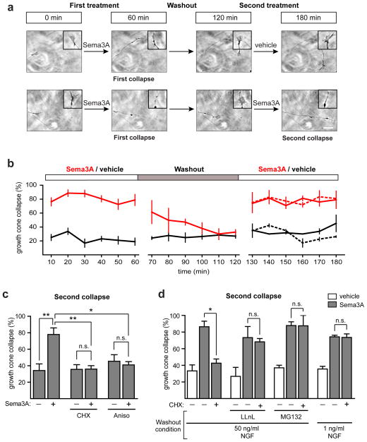
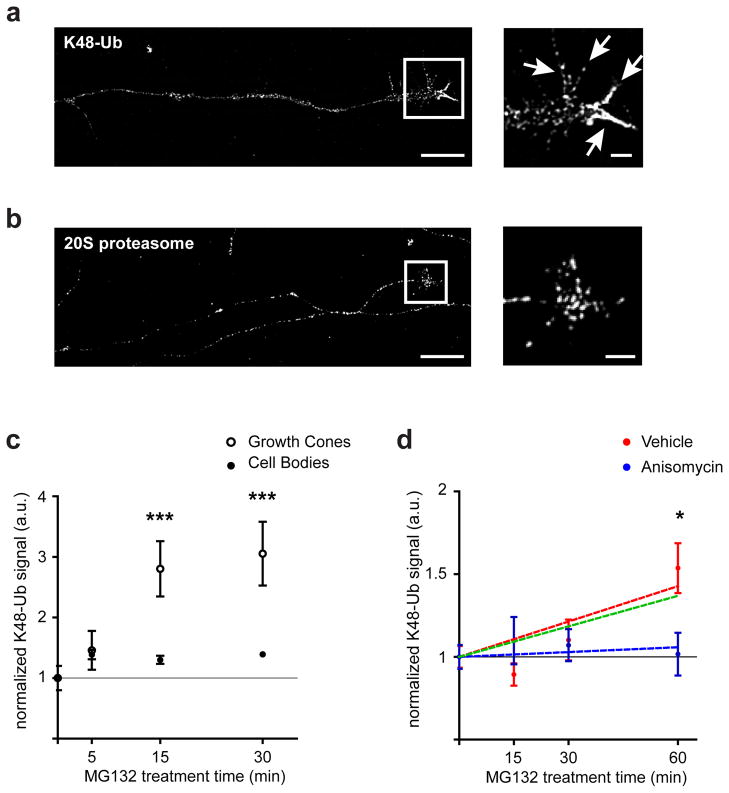
Local translation mediates axonal responses to Semaphorin3A (Sema3A) and other guidance cues. However, only a subset of the axonal proteome is locally synthesized, whereas most proteins are trafficked from the soma. The reason why only specific proteins are locally synthesized is unknown. Here we show that local protein synthesis and degradation are linked events in growth cones. We find that growth cones exhibit high levels of ubiquitination and that local signalling pathways trigger the ubiquitination and degradation of RhoA, a mediator of Sema3A-induced growth cone collapse. Inhibition of RhoA degradation is sufficient to remove the protein-synthesis requirement for Sema3A-induced growth cone collapse. In addition to RhoA, we find that locally translated proteins are the main targets of the ubiquitin-proteasome system in growth cones. Thus, local protein degradation is a major feature of growth cones and creates a requirement for local translation to replenish proteins needed to maintain growth cone responses.


Conflict of interest statement

COMPETING INTERESTS

The authors declare no competing financial interests.

Figures

Figure 1
Figure 1. The UPS and NGF maintain the local protein synthesis dependence of Sema3A-induced growth cone collapse
(a) Growth cones can undergo sequential rounds of Sema3A-induced growth cone collapse. Representative phase images of the same growth cones at t=0 min (before any treatment), t=60 min (after 1 h of Sema3A or vehicle exposure), t=120 min (after washout) and t=180 min (after 1 h of the second Sema3A or vehicle treatment). Scale bar represents 5 μm. The insets show magnified images of the growth cones. (b) Quantification of results in (a). The red line represents growth cones exposed to 450 ng per ml Sema3A while the black line represents vehicle-treated growth cones. In the last hour of the experiment (right side), the dashed red line indicates growth cones that were treated with vehicle in the first hour and Sema3A in the final hour, while the solid red line represents growth cones that had been exposed to Sema3A in the first hour and were again exposed to Sema3A in the final hour of the experiment. Growth cones treated with vehicle during the final hour are indicated with a black line (dashed for vehicle-treated during the first hour, solid for Sema3A-treated during the first hour). n≥15 growth cones per time point from at least three independent experiments. (c) The second Sema3A-induced growth cone collapse is dependent on local protein synthesis. Treatment of growth cones with the protein synthesis inhibitors cycloheximide (CHX, 10 μM) or anisomycin (Aniso, 40 μM) abolished the second Sema3A-induced growth cone collapse. **p<0.01, one-way ANOVA with Turkey’s multiple comparison test, n≥38 growth cones per condition from at least three independent experiments. (d) The requirement of local protein synthesis in Sema3A-induced growth cone collapse depends on UPS activity and NGF levels during washout. Addition of proteasome inhibitors (50 μM LLnL or 5 μM MG132) during the washout phase or washout in media containing minimal NGF concentrations (1 ng per ml) rendered the second collapse protein synthesis independent. *p<0.05, one-way ANOVA with Turkey’s multiple comparison test, n≥34 growth cones per condition from at least three independent experiments.
Figure 2
Figure 2. NGF signaling induces local recruitment of the UPS via Smurf1
(a,b) NGF-dependent axon outgrowth requires the UPS. Axonal application of NGF leads to a significant increase in the rate of axon outgrowth that is abolished by treatment of axons with the proteasome inhibitors LLnL (50 μM) or lactacystin (10 μM). Phase images show representative axons at t=0 and t=60 min after exposure to no NGF (a) or 50 ng per ml NGF (b). The yellow triangles indicate the position of the growth cone at the beginning of the experiment (t=0 min) while the blue triangles indicate the position of the growth cone at the end of the experiment (t=60 min). Scale bar represents 10 μm. (c) Quantification of results in (A–B). Axon growth was calculated by measuring the distance travelled by the growth cone between t=0 and t=60 min. **p<0.01, one-way ANOVA with Turkey’s multiple comparison test, n≥71 axons per condition from at least three independent experiments. (d) Smurf1 levels and localization within growth cones change upon NGF treatment. DIV3-4 rat DRG neurons were exposed to 0 or 50 ng/ml NGF for 10 min, followed by fixation and staining. Immunofluorescence images of endogenous Smurf1 (green) and Alexa-568-phalloidin (red) show that exposure to NGF increases the amount of Smurf1 in growth cones, with more Smurf1 localizing to filopodia (white arrows). (e) Smurf1 is required for NGF-induced axon outgrowth. NGF-induced axon outgrowth is abolished in Smurf1 knockdown cells compared to wild-type or shLacZ controls. The basal growth rate is not affected by Smurf1 knockdown. **p<0.01, ***p<0.001, one-way ANOVA with Turkey’s multiple comparison tests, n≥87 axons per condition from at least three independent experiments.
Figure 3
Figure 3. Smurf1 maintains the protein synthesis requirement in Sema3A-induced growth cone collapse
(a) Smurf1 accounts for the protein synthesis dependence of Sema3A-induced growth cone collapse. Rat DRG neurons were transduced upon plating with lentiviral constructs expressing two distinct Smurf1 shRNAs or a control non-targeting shRNA (shLacZ) and grown for 5 DIV. Application of anisomycin (Aniso, 40 μM) reduced Sema3A-induced growth cone collapse in neurons expressing shLacZ but not in Smurf1 knockdown neurons. **p<0.01, one-way ANOVA with Turkey’s multiple comparison test, n≥58 growth cones per condition from at least three independent experiments. (b) Overexpression of a catalytically inactive mutant form of Smurf1 (Smurf1 C699A) leads to a Sema3A-induced growth cone collapse that is protein synthesis independent. *p<0.05, **p<0.01, one-way ANOVA with Turkey’s multiple comparison test, n≥53 growth cones per condition from at least three independent experiments.
Figure 4
Figure 4. Smurf1 degradation of RhoA leads to the protein synthesis requirement in Sema3A-induced growth cone collapse
(a) NGF induces rapid ubiquitination of endogenous RhoA in PC12 cells. PC12 cells were exposed to NGF (100 ng per ml) for the indicated times. Endogenous RhoA was immunoprecipitated with an anti-RhoA antibody and the degree of ubiquitination was evaluated by Western Blot with an anti-ubiquitin antibody. (b) NGF induces proteasomal-mediated degradation of endogenous RhoA in growth cones of DRG neurons. Neurons were incubated in minimal NGF (1 ng per ml) for 18 h to decrease NGF signaling. DRG neurons were then exposed to either 0 or 50 ng per ml NGF either in the presence (NGF + MG132) or absence of MG132 (5 μM) for the indicated times, and fixed and stained to quantify the levels of endogenous RhoA within growth cones. Scale bar represents 10 μm. (c) Quantification of results in (b). RhoA levels were normalized to the levels at 0 min (dotted line). For quantification purposes, the RhoA immunofluorescence signal was normalized to that of GAP43. *p<0.05, one-way ANOVA, n≥35 per condition from at least three independent experiments. (d) Smurf1 induces degradation of endogenous RhoA. RhoA levels were determined in untransfected cells (−) and in cells expressing wild type Smurf1 (WT) or a catalytically inactive mutant form of Smurf1 (Smurf1 C699A).
Figure 5
Figure 5. Smurf1 ubiquitinates RhoA at Lys 6, 7, and 51
(a) RhoA is degraded by Smurf1 through ubiquitination of Lys 6, 7 and 51. Flag-tagged wild type or mutant RhoA-expressing plasmids were co-transfected with GFP- or Smurf1-expressing plasmids in HEK293T cells using calcium phosphate transfection. (b) RhoA Lys 6, 7, and 51 are in close proximity in the crystal structure. The three-dimensional structure of RhoA (PDB: 2A3B) showed that Lys 51 is positioned near Lys 6 and 7 (black arrows) on the same surface. Lysines are indicated in magenta while the rest of the protein is in blue. (c) RhoA degradation is required to maintain the protein synthesis dependence of Sema3A-induced growth cone collapse. Sema3A-induced growth cone collapse was evaluated in neurons expressing GST (GST), wild type RhoA (RhoA) or the K6/7/51R mutant form of RhoA (K6/7/51R). The samples were treated with Sema3A (450 ng per ml) and with DMSO or anisomycin (40 μM). *p<0.05, one-way ANOVA with Turkey’s multiple comparison test, n≥51 growth cones per condition from at least three independent experiments.
Figure 6
Figure 6. Growth cones exhibit a high degree of ubiquitination and protein turnover
(a) Growth cones display high levels of ubiquitination. DRG neurons cultured in standard growth conditions were immunostained with K48-linkage specific polyubiquitin antibody (K48-Ub). The white arrows in the inset indicate the filopodia. Scale bar represents 10 μm, inset scale bar represents 2 μm. (b) Growth cones are enriched in 20S proteasomal subunits, which primarily localize to the palm region of growth cones. DRG neurons were immunostained with an antibody recognizing the 20S proteasome. Scale bar represents 10 μm, inset scale bar represents 2 μm. (c) Growth cone proteins are rapidly ubiquitinated. DRG neurons were treated with MG132 (5 μM) for the times indicated and stained for K48-Ub. The signal was normalized for volume by tau-1 staining and measured in growth cones (open circles) and cell bodies (closed circles. ***p<0.001, Student’s t-test, n≥27 growth cones and cell bodies per condition per time point from at least three independent experiments. (d) Locally synthesized proteins are the primary target of ubiquitination in growth cones. DIV5 DRG axons were treated for the indicated times with MG132 and stained for K48-Ub. The immunofluorescence signal in growth cones was normalized to the average signal for vehicle-treated controls. Red and blue circles indicates treated with vehicle or anisomycin (40 μM), respectively. The ubiquitination of the locally translated pool of proteins (green line) can be extrapolated by subtracting the ubiquitination rate of the cell body-derived pool (blue line, linear regression) from the total axonal protein pool (red line, linear regression). The locally synthesized protein pool accounts for the majority of the ubiquitination seen in axonal growth cones. *p<0.05, Student’s t-test, n≥26 growth cones per condition from at least three independent experiments.

Similar articles

Cited by

References

    1. Zivraj KH, et al. Subcellular profiling reveals distinct and developmentally regulated repertoire of growth cone mRNAs. J Neurosci. 2010;30:15464–15478. - PMC - PubMed
    1. Jung H, Yoon BC, Holt CE. Axonal mRNA localization and local protein synthesis in nervous system assembly, maintenance and repair. Nat Rev Neurosci. 2012;13:308–324. - PMC - PubMed
    1. Deglincerti A, Jaffrey SR. Insights into the roles of local translation from the axonal transcriptome. Open Biology. 2012;2:120079–120089. - PMC - PubMed
    1. Campbell DS, Holt CE. Chemotropic responses of retinal growth cones mediated by rapid local protein synthesis and degradation. Neuron. 2001;32:1013–1026. - PubMed
    1. Wu KY, et al. Local translation of RhoA regulates growth cone collapse. Nature. 2005;436:1020–1024. - PMC - PubMed

Publication types

MeSH terms

LinkOut - more resources