. 2015 Apr 22;11(4):e1005039.
doi: 10.1371/journal.pgen.1005039. eCollection 2015 Apr.

Re-replication of a centromere induces chromosomal instability and aneuploidy

Affiliations

Re-replication of a centromere induces chromosomal instability and aneuploidy

Stacey L Hanlon et al. PLoS Genet. .

Abstract

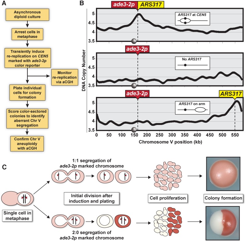
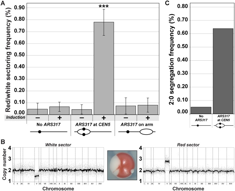
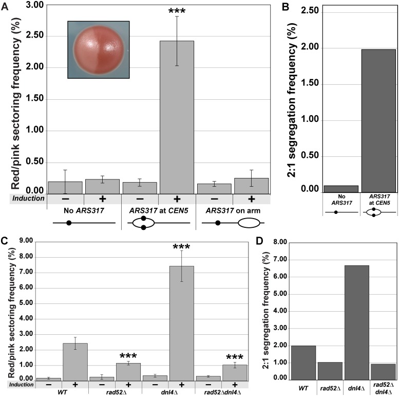
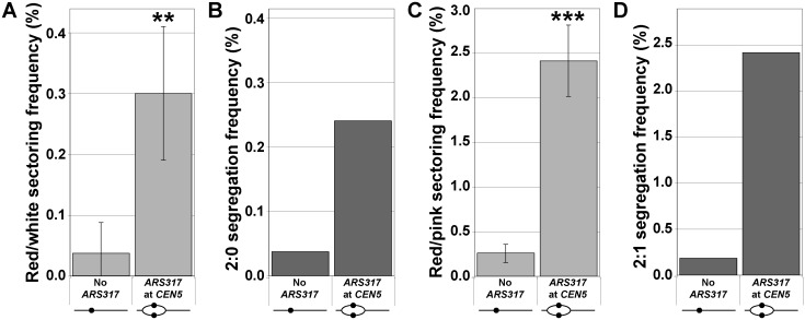
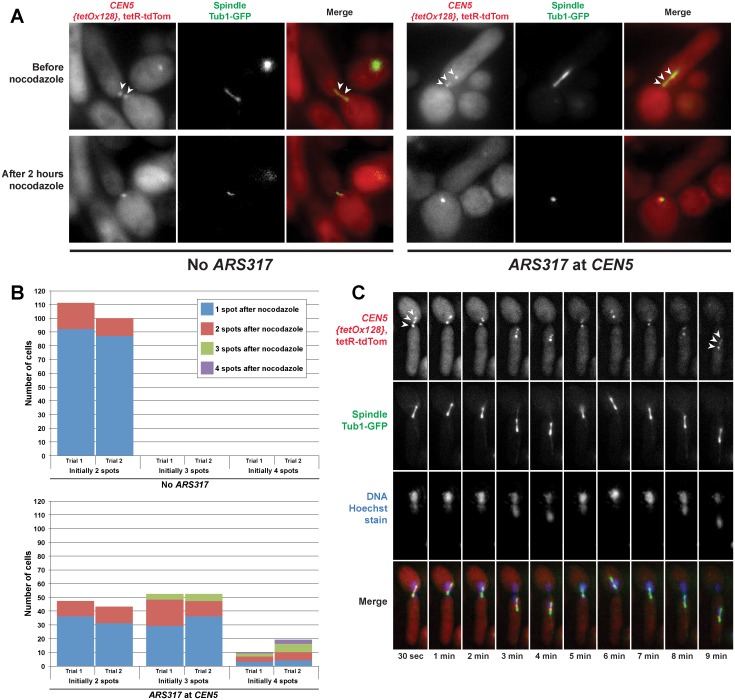
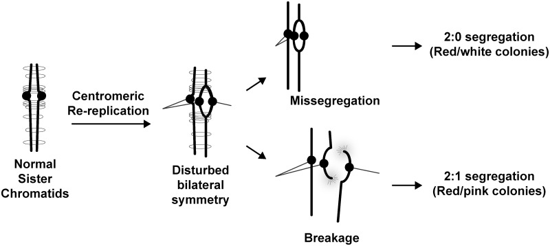
The faithful inheritance of chromosomes during cell division requires their precise replication and segregation. Numerous mechanisms ensure that each of these fundamental cell cycle events is performed with a high degree of fidelity. The fidelity of chromosomal replication is maintained in part by re-replication controls that ensure there are no more than two copies of every genomic segment to distribute to the two daughter cells. This control is enforced by inhibiting replication initiation proteins from reinitiating replication origins within a single cell cycle. Here we show in Saccharomyces cerevisiae that re-replication control is important for the fidelity of chromosome segregation. In particular, we demonstrate that transient re-replication of centromeric DNA due to disruption of re-replication control greatly induces aneuploidy of the re-replicated chromosome. Some of this aneuploidy arises from missegregation of both sister chromatids to one daughter cell. Aneuploidy can also arise from the generation of an extra sister chromatid via homologous recombination, suggesting that centromeric re-replication can trigger breakage and repair events that expand chromosome number without causing chromosomal rearrangements. Thus, we have identified a potential new non-mitotic source of aneuploidy that can arise from a defect in re-replication control. Given the emerging connections between the deregulation of replication initiation proteins and oncogenesis, this finding may be relevant to the aneuploidy that is prevalent in cancer.


Conflict of interest statement

The authors have declared that no competing interests exist.

Figures

Fig 1
Fig 1. Monitoring chromosome segregation fidelity after centromeric re-replication.
(A) Experimental flowchart starting with diploid re-replicating cells containing one Chromosome V homolog marked with the ade3–2p copy number reporter. (B) Re-replication profile of Chromosome V for diploid cells arrested in metaphase (with baseline copy number of 4C) and induced to re-replicate (see S1 Table). ARS317 and ade3–2p mark integration sites of the preferentially reinitiating origin and the copy number reporter, respectively. Inset shows schematic of re-replication bubbles inferred from profiles. Circles on X-axis and in schematic represent centromere CEN5. (C) Illustration showing how 1:1 segregation of the ade3–2p marked homolog in the first cell division after centromere re-replication leads to pink colonies and 2:0 missegregation leads to red/white sectored colonies.
Fig 2
Fig 2. Centromeric re-replication causes 2:0 missegregation of chromosomes.
(A) Centromeric re-replication induces red/white sectored colonies. Diploid re-replicating strains (characterized in Fig. 1B and induced to re-replicate as described in Fig. 1A) were scored for the frequency of red/white sectored colonies either before (-) or after (+) induction of re-replication (see S7 Table). Frequencies are presented as the mean ± SD (n ≥ 3). When compared to no ARS317 at CEN5, the frequency after re-replication was significantly different for ARS317 at CEN5 (***, p = 6.03x10⁻⁸) but not for ARS317 on the right arm (p = 0.175). (B) aCGH copy number analysis of a representative red/white colony that was scored as a 2:0 segregation event (see Materials and Methods). (C) Estimated frequency of 2:0 segregation events after 3 hr of re-replication. The average sectoring frequency for each strain shown in (A) was multiplied by the fraction of aCGH-analyzed isolates that showed 2:0 segregation of the ade2–3p marked Chromosome V homolog (see S2 and S8 Tables).
Fig 3
Fig 3. Centromeric re-replication causes 2:1 segregation through chromosome gain.
(A) Centromeric re-replication induces red/pink sectored colonies. Diploid re-replicating strains characterized in Fig. 1B and induced to re-replicate as described in Fig. 1A, were scored for the frequency of red/pink sectored colonies (see inset) either before (-) or after (+) induction of re-replication (see S7 Table). Frequencies are presented as the mean ± SD (n ≥ 3). When compared to no ARS317 at CEN5, the frequency after re-replication was significantly different for ARS317 at CEN5 (***, p = 3.3x10⁻⁸) but not for ARS317 on the right arm (p = 0.253). (B) Estimated frequency of 2:1 segregation events after 3 hr of re-replication. The average sectoring frequency for each strain shown in (A) was multiplied by the fraction of aCGH-analyzed isolates that showed 2:1 segregation of the ade2–3p marked Chromosome V homolog (see S3 and S8 Tables). (C) Dependence of red/pink colony frequencies induced by centromeric re-replication on recombination. Diploid re-replicating strains with reinitiating origin ARS317 at CEN5 and homozygous deletions of the indicated genes were scored for the frequency of red/pink sectored colonies as described and presented as in (A) (see S7 Table). When compared to the undeleted WT background, the frequency after re-replication was significantly different for rad52∆ (***, p = 5.8x10⁻⁶), dnl4∆ (***, p = 2.1x10⁻⁹), and rad52∆ dnl4∆ (***, p = 7.4x10⁻⁵). (D) Dependence of 2:1 segregation events induced by centromeric re-replication on homologous recombination. Segregation events were estimated as described in (B) using the frequencies reported in (C) (see S3 and S8 Tables).
Fig 4
Fig 4. Centromeric re-replication induced in cycling cells causes 2:0 and 2:1 segregation.
Re-replication was induced for 3 hr in unarrested cycling cells (see S4 Fig), which were then analyzed as described in Fig. 1A. (A) Frequencies of red/white sectored colonies after re-replication (see S7 Table) are presented as the mean ± SD (n ≥ 3) and shown to be significantly higher for ARS317 at CEN5 versus no ARS317 at CEN5 (** p = 0.002). (B) Estimates of 2:0 segregation frequencies after re-replication were calculated as described for Fig. 2C (see S4 and S8 Tables). (C) Frequencies of red/pink sectored colonies after re-replication (see S7 Table) are presented as the mean ± SD (n ≥ 3) and shown to be significantly higher for ARS317 at CEN5 versus no ARS317 at CEN5 (*** p = 2.2x10⁻⁵). (D) Estimates of 2:1 segregation frequencies after re-replication were calculated as described for Fig. 3B (see S5 and S8 Tables).
Fig 5
Fig 5. Spindle dependent dynamic separation and movement of re-replicated centromeres.
Exponentially-growing cells with either ARS317 integrated near CEN5 (YJL10671/YJL10672) or not integrated at all (YJL10665/YJL10666) were induced to re-replicate for three hours by addition of galactose. During this time, the DNA damage response triggered by re-replication caused both strains to arrest in metaphase with intact mitotic spindles. Cells were shifted to dextrose containing media to limit further induction of re-replication before being imaged live, initially in the absence of nocodazole then 2 hr after addition of nocodazole. (A) Spots corresponding to the TET operator arrays positioned near CEN5 and bound to tdTomato-tagged TET repressors are indicated by arrowheads. (B) Quantification of the number of spots observed before and after nocodazole addition in cells with ARS317 near CEN5 or without ARS317. The number of cells scored pre-nocodazole is charted based on initial spot number, with each bar divided into the number of cells retaining one, two, three, or four spots after nocodazole treatment (see S6 Table). Each strain in both trials was scored for ≥ 100 cells. (C) Video microscopy in a single Z-plane of a live cell that has undergone centromeric re-replication. The three spots corresponding to TET operator arrays bound to tdTomato-tagged TET repressors (on the left of CEN5) are indicated by arrowheads in the first and last panels.
Fig 6
Fig 6. Possible ways for centromeric re-replication to perturb chromosome segregation.
Normal sister chromatids are bilaterally symmetric and held together via cohesin to ensure their bi-orientation with respect to the spindle poles. Centromere re-replication disrupts this bilateral symmetry and can lead to abnormal bipolar attachment of a single chromatid to both spindle poles. During anaphase, this bipolar attachment could lead to a 2:0 segregation pattern. Alternatively the affected sister chromatid could break and repair in a RAD52-dependent manner to produce a 2:1 segregation pattern. Also conceivable but not shown are disruption of kinetochore function or pericentromeric cohesion by re-replication.

Similar articles

Cited by

References

    1. Arias EE, Walter JC (2007) Strength in numbers: preventing rereplication via multiple mechanisms in eukaryotic cells. Genes Dev 21: 497–518. - PubMed
    1. Hook SS, Lin JJ, Dutta A (2007) Mechanisms to control rereplication and implications for cancer. Curr Opin Cell Biol 19: 663–671. - PMC - PubMed
    1. Siddiqui K, On KF, Diffley JFX (2013) Regulating DNA Replication in Eukarya. Cold Spring Harb Perspect Biol. - PMC - PubMed
    1. Green BM, Morreale RJ, Ozaydin B, Derisi JL, Li JJ (2006) Genome-wide mapping of DNA synthesis in Saccharomyces cerevisiae reveals that mechanisms preventing reinitiation of DNA replication are not redundant. Mol Biol Cell 17: 2401–2414. - PMC - PubMed
    1. Wilmes GM, Archambault V, Austin RJ, Jacobson MD, Bell SP, et al. (2004) Interaction of the S-phase cyclin Clb5 with an “RXL” docking sequence in the initiator protein Orc6 provides an origin-localized replication control switch. Genes Dev 18: 981–991. - PMC - PubMed

Publication types

Associated data