. 2015 Apr 22:6:6898.
doi: 10.1038/ncomms7898.

Regulation of endogenous transmembrane receptors through optogenetic Cry2 clustering

Affiliations

Regulation of endogenous transmembrane receptors through optogenetic Cry2 clustering

L J Bugaj et al. Nat Commun. .

Abstract

Transmembrane receptors are the predominant conduit through which cells sense and transduce extracellular information into intracellular biochemical signals. Current methods to control and study receptor function, however, suffer from poor resolution in space and time and often employ receptor overexpression, which can introduce experimental artefacts. We report a genetically encoded approach, termed Clustering Indirectly using Cryptochrome 2 (CLICR), for spatiotemporal control over endogenous transmembrane receptor activation, enabled through the optical regulation of target receptor clustering and downstream signalling using noncovalent interactions with engineered Arabidopsis Cryptochrome 2 (Cry2). CLICR offers a modular platform to enable photocontrol of the clustering of diverse transmembrane receptors including fibroblast growth factor receptor (FGFR), platelet-derived growth factor receptor (PDGFR) and integrins in multiple cell types including neural stem cells. Furthermore, light-inducible manipulation of endogenous receptor tyrosine kinase (RTK) activity can modulate cell polarity and establish phototaxis in fibroblasts. The resulting spatiotemporal control over cellular signalling represents a powerful new optogenetic framework for investigating and controlling cell function and fate.


Conflict of interest statement

Competing financial interests: The authors declare no competing financial interests.

Figures

Figure 1
Figure 1
The CLICR strategy enables Cry2 activation of transmembrane receptors. (a) CLICR allows modular Cry2 clustering and activation of membrane receptors via non-covalent interactions, avoiding complications associated with overexpression of receptor fusions. With CLICR, Cry2 fused to a receptor-targeting binding domain (BD) is expressed in the cytoplasm. In the dark, unclustered state, BD affinity for the receptor is weak and imparts no or little membrane localization. Upon light induced clustering, BD-Cry2 oligomers increase local BD concentration, enabling membrane translocation, binding, and nucleation of a receptor cluster. (b) CLICR clustering allows activation of cytoplasmic protein targets. Upon light activation, the Cry2-mCh-LZa (red) and LZb-LRP6c-GFP (green) components co-cluster (c), suggesting successful clustering of non-covalently bound proteins via homo-oligomerization of Cry2. (d,e) CLICR clustering also enables membrane translocation of the initially cytoplasmic Cry2-mCh-LZa. Scale bars = 20 μm.
Figure 2
Figure 2
CLICR can target and cluster endogenous transmembrane receptors. (a) To target endogenous receptors with CLICR, an adapter domain with affinity for native receptors was used in place of the engineered LZa-LZb interaction used previously. The SH2 domain from PLCγ was used as a binding domain fused to the N-terminus of mCh-Cry2 (SH2-N) to target endogenous receptor tyrosine kinases. Illuminated SH2-N expressed in 293Ts (b, top panels) localized to membrane-anchored foci. In 3T3 fibroblasts (c, top panels), illumination of SH2-N induced translocation to the membrane periphery and to structures resembling focal adhesions. In neural stem cells (NSC) (d, top panels), illuminated SH2-N localized to immobilized membrane clusters both in the cell body and in neurites. In all 3 cell types, inhibition with an FGFR1/PDGFRβ/EGFR-specific inhibitor (RTKi) largely abrogated visible SH2-N translocation. (b, c, d, lower panels). In 293Ts, upon blue light illumination SH2-N colocalized (arrows) to exogenously expressed, full-length FGFR1 (e) and PDGFRβ (g) fused to GFP, and this colocalization was abrogated in the presence of inhibitors specific to these receptors (f, h). Scale bars = 20 μm. Light-stimulated association of SH2-N with either FGFR1-GFP or PDGFRβ-GFP was confirmed through co-immunoprecipitation of SH2-N with the receptors (i).
Figure 3
Figure 3
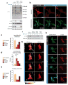
CLICR clustering allows photoactivation of endogenous RTKs in fibroblasts and enables PDGFRβ-dependent phototaxis. Western blot analysis (a) of serum-starved fibroblasts treated for 10 minutes with blue light shows broad upregulation of tyrosine phosphorylation in illuminated vs unilluminated samples, and this difference is abrogated upon 10 μm RTK inhibition. Samples grown in 2% serum were used as a positive control. In particular, light-treated samples demonstrated an RTK-dependent increase in Akt and Erk1/2 phosphorylation. (b) Time lapse imaging of SH2-N expressing fibroblasts shows lamellipodial extension and cell repolarization upon whole field blue light exposure. (c) The proportion of cells extending lamellipodia in response to whole field illumination was reduced by broad RTK or PDGFR inhibition, but not FGFR inhibition. Error bars represent 95% confidence intervals, n = 24–83 cells per condition. (d) Among cells extending light-induced lamellipodia under broad RTK inhibition or PDGFR inhibition, a longer delay before initial lamellipodial extension was evident, while FGFR inhibition did not induce a delay in extension. Graph shows means ± 1 s.d, n = 4–58 cells per condition. ***p < 0.001 by one way ANOVA with Tukey pairwise analysis. (e) The proportion of cells exhibiting light-induced polarization was reduced by RTKi or PDGFRi treatment, and slightly reduced by FGFRi treatment. Error bars represent 95% confidence intervals, n = 24–83 cells per condition. (f) Focal illumination enabled the spatial definition of the lamellipodial extension region coupled with re-establishment of a PIP3 gradient at the site of illumination (arrows), as observed through a PH(Akt)-Venus biosensor. (g) CLICR-enabled focal activation of lamellipodial extension and polarity establishment allows the effective rewiring of PDGFR-dependent chemotaxis to respond to light. Fibroblasts co-expressing SH2-N and Lifeact-Venus were illuminated focally, and actin polymerization, lamellipodial extension, and cell motility were induced in the direction of the illuminated region. Illuminating the trailing edge induced re-polarization and a reversal of migration direction. Scale bars = 20 μm.
Figure 4
Figure 4
CLICR targeting and clustering of β-integrins (a) Co-immunoprecipitation of the talin F3 domain fused to mCh-Cry2 (F3-N) co-expressed with integrin-β3-GFP in HEK 293T cells reveals strong association of the constructs under blue light exposure. (b) Fibroblasts expressing F3-N show light-induced formation of puncta that move within the cell along defined linear trajectories (see Supplementary Movie 9). (c) Co-expression of F3-N and integrin-β3-GFP in 3T3 cells demonstrates a high degree of co-clustering of the two constructs under blue light, with reversion of clusters to a diffuse state in the dark. (d) All co-expressing cells in (c) that displayed F3-N clustering exhibited concomitant β3-integrin co-clustering. Further analysis of quantifiable cellular regions (e) revealed that all observable F3-N clusters co-clustered with integrin-β3-GFP (f). Scale bars = 20 μm.

Similar articles

Cited by

References

    1. Coward P, et al. Controlling signaling with a specifically designed Gi-coupled-receptor. Proceedings of the National Academy of Sciences. 1998;95:352–357. - PMC - PubMed
    1. Spencer D, Wandless T, Schreiber S, Crabtree G. Controlling signal transduction with synthetic ligands. Science. 1993;262:1019–1024. - PubMed
    1. Dikic I, Schlessinger J, Lax I. PC12 cells overexpressing the insulin receptor undergo insulin-dependent neuronal differentiation. Current Biology. 1994;4:702–708. - PubMed
    1. Jiang Y, Woronicz JD, Liu W, Goeddel DV. Prevention of Constitutive TNF Receptor 1 Signaling by Silencer of Death Domains. Science. 1999;283:543–546. - PubMed
    1. Traverse S, et al. EGF triggers neuronal differentiation of PC12 cells that overexpress the EGF receptor. Current Biology. 1994;4:694–701. - PubMed

Publication types