. 2015 Apr 22;10(4):e0125206.
doi: 10.1371/journal.pone.0125206. eCollection 2015.

Increase in Red Blood Cell-Nitric Oxide Synthase Dependent Nitric Oxide Production during Red Blood Cell Aging in Health and Disease: A Study on Age Dependent Changes of Rheologic and Enzymatic Properties in Red Blood Cells

Affiliations

Increase in Red Blood Cell-Nitric Oxide Synthase Dependent Nitric Oxide Production during Red Blood Cell Aging in Health and Disease: A Study on Age Dependent Changes of Rheologic and Enzymatic Properties in Red Blood Cells

Daniel Alexander Bizjak et al. PLoS One. .

Abstract

Aim: To investigate RBC-NOS dependent NO signaling during in vivo RBC aging in health and disease.

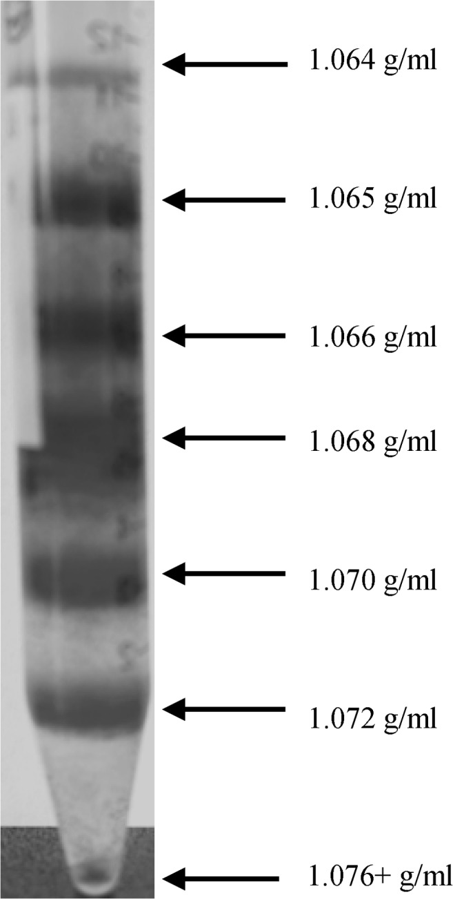
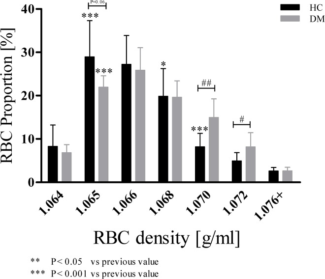
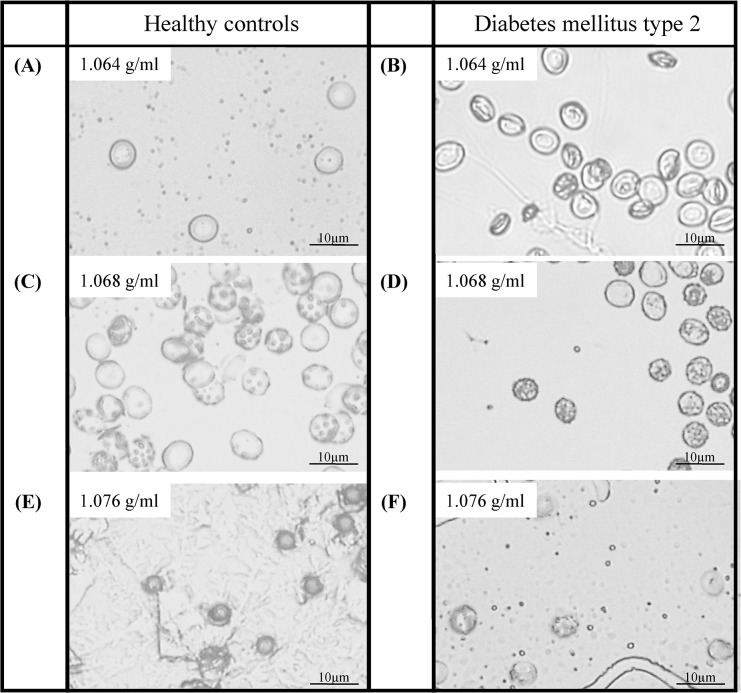
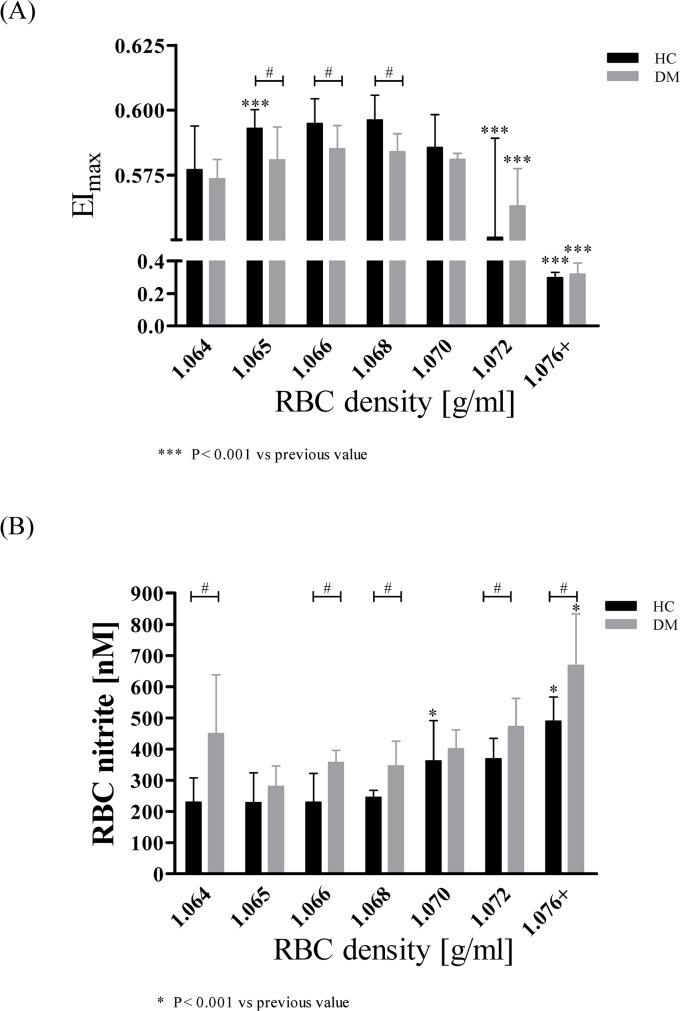
Method: RBC from fifteen healthy volunteers (HC) and four patients with type 2 diabetes mellitus (DM) were separated in seven subpopulations by Percoll density gradient centrifugation.

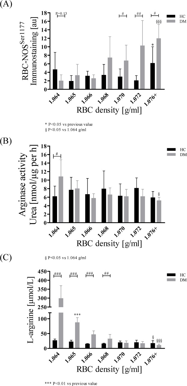
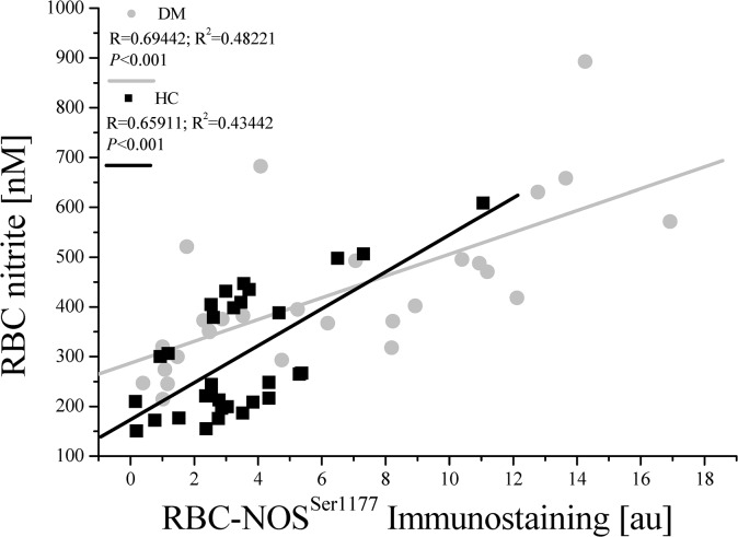
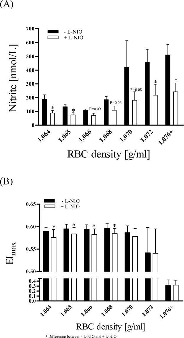
Results: The proportion of old RBC was significantly higher in DM compared to HC. In both groups, in vivo aging was marked by changes in RBC shape and decreased cell volume. RBC nitrite, as marker for NO, was higher in DM and increased in both HC and DM during aging. RBC deformability was lower in DM and significantly decreased in old compared to young RBC in both HC and DM. RBC-NOS Serine1177 phosphorylation, indicating enzyme activation, increased during aging in both HC and DM. Arginase I activity remained unchanged during aging in HC. In DM, arginase I activity was significantly higher in young RBC compared to HC but decreased during aging. In HC, concentration of L-arginine, the substrate of RBC-NOS and arginase I, significantly dropped from young to old RBC. In DM, L-arginine concentration was significantly higher in young RBC compared to HC and significantly decreased during aging. In blood from healthy subjects, RBC-NOS activation was additionally inhibited by N5-(1-iminoethyl)-L-Ornithine dihydrochloride which decreased RBC nitrite, and impaired RBC deformability of all but the oldest RBC subpopulation.

Conclusion: This study first-time showed highest RBC-NOS activation and NO production in old RBC, possibly to counteract the negative impact of cell shrinkage on RBC deformability. This was even more pronounced in DM. It is further suggested that highly produced NO only insufficiently affects cell function of old RBC maybe because of isolated RBC-NOS in old RBC thus decreasing NO bioavailability. Thus, increasing NO availability may improve RBC function and may extend cell life span in old RBC.


Conflict of interest statement

Competing Interests: The authors have declared that no competing interests exist.

Figures

Fig 1
Fig 1. Red blood cell layers after Percoll density gradient centrifugation.
Seven RBC fractions were generated by Percoll density gradient centrifugation. Least dense fraction on the top consisted of youngest RBC; increasing density indicated increasing RBC age. Arrows indicate the respective density of Percoll solution. Subpopulations were clearly separated and collected. Oldest fraction at the bottom was described as 1.076+ g/ml, with “+” indicating possible even denser RBC than the densest Percoll layer of 1.076 g/ml.
Fig 2
Fig 2. RBC proportion of HC and DM in dependence of increased cell age.
Bars show RBC proportion of fractionated subpopulations to total RBC volume, which were divided into young (1.064 g/ml), middle old (1.065–1.068 g/ml) and old (1.070–1.076+ g/ml) RBC. In both study groups, middle old RBC represented the main proportion on total RBC volume. DM showed significantly higher amount of old RBC compared to HC. Data are presented as mean ± standard deviation of n = 15 (HC) and n = 4 (DM).
Fig 3
Fig 3. Representative microscopic pictures of the seven isolated RBC subpopulations.
Density of the RBC increased with increasing cell age, and cell shape changed from discoid to echinocyte. In RBC of DM, cell shape changes were more pronounced. Magnification for all pictures was 400-fold.
Fig 4
Fig 4. Mean Cellular Volume (MCV) and phosphatidylserine (PS) externalisation with increasing cell age in HC and DM.
(A) Mean cellular volume constantly decreased during aging in both groups. MCV of young and middle aged RBC was significantly decreased in DM compared to HC. RBC of HC lost 20% of MCV during aging (P < 0.001; comparison of old and young RBC). RBC of DM lost 12% of MCV during aging (P < 0.05; comparison of old and young RBC). Data are presented as mean ± standard deviation of n = 15 (HC) and n = 4 (DM). (B) In both groups, Annexin V, a marker for PS externalisation, significantly increased in the oldest subpopulation (P < 0.05 compared to 1.072 g/ml). Data are presented as mean ± standard deviation of n = 4 (HC) and n = 4 (DM).
Fig 5
Fig 5. Maximal RBC deformability (EImax) and nitrite concentration in RBC of HC and DM during cell aging.
(A) In RBC of HC, EImax values increased from 1.064 g/ml to 1.065 g/ml (P < 0.001), remained constant to 1.068 g/ml and then decreased with increasing cell age. Oldest RBC (1.076+ g/ml) showed lowest EImax (P < 0.001 compared to 1.072 g/ml). EImax of middle aged RBC (1.065–1.068 g/ml) was significantly lower in DM compared to HC. EImax of DM significantly decreased during aging. Data are presented as mean ± standard deviation of n = 15 (HC) and n = 4 (DM). (B) In HC, RBC nitrite concentration remained constant from 1.064 g/ml to 1.068 g/ml and then significantly increased reaching its maximum values at 1.076+ g/ml. RBC nitrite concentration was significantly higher in DM compared to HC for 1.064, 1.066, 1.0681.072 and 1.076+ g/ml. RBC nitrite concentration in RBC of DM increased during aging. Data are presented as mean ± standard deviation of n = 5 (HC) and n = 4 (DM).
Fig 6
Fig 6. RBC-NOS activation, arginase I activity and L-arginine concentration in RBC of HC and DM during increasing cell age.
(A) In HC, RBC-NOS phosphorylation at its Serine1177 residue, representing activation of the enzyme, was significantly increased in subpopulation 1.076+ g/ml (P < 0.05 compared to 1.072 g/ml). In DM, RBC-NOSSer1177 significantly increased during aging with significantly higher values obtained in DM compared to HC in RBC fractions 1.070, 1.072 and 1.076+ g/ml. Data are presented as mean ± standard deviation of n = 8 (HC) and n = 4 (DM). (B) Arginase I activity was represented by the synthesis of the arginase I product urea. In both groups, arginase activity remained constant in all measured subpopulations. Data are presented as mean ± standard deviation of n = 4 (HC) and n = 4 (DM). (C) In HC, L-arginine concentration remained unchanged during aging. DM showed significantly higher L-arginine values compared to HC for young and middle old RBC (1.064–1.068 g/ml). L-arginine concentration of DM decreased with increasing cell age. Data are presented as mean ± standard deviation of n = 8 (HC) and n = 4 (DM).
Fig 7
Fig 7. Linear regression of RBC-NOSSer1177 immunostaining and RBC nitrite concentration in HC and DM.
Calculation of R and R2 (goodness of fit) revealed positive correlation of RBC-NOSSer1177 and RBC nitrite for both study groups.
Fig 8
Fig 8. Nitrite concentration and maximum RBC deformability (EImax) in RBC after L-NIO incubation in comparison to untreated RBC.
(A) RBC nitrite concentration was decreased in all subpopulations by L-NIO. Data are presented as Mean ± Standard Error of MEAN (n = 10). (B) EImax values significantly decreased in subpopulations of 1.064 g/ml to 1.068 g/ml after L-NIO incubation. No decrease was observed in old subpopulations (1.070 g/ml and 1.076+ g/ml). Data are presented as Mean ± Standard Deviation (n = 10).

References

    1. Palis J. Primitive and definitive erythropoiesis in mammals. Front in Physio. 2014. January 28; 5:3. - PMC - PubMed
    1. Franco RS. The measurement and importance of red cell survival. Am J Hematol. 2009;84(2): 109–14. 10.1002/ajh.21298 - DOI - PubMed
    1. Rifkind JM, Nagababu E. Hemoglobin Redox Reactions and Red Blood Cell Aging. Antioxid Redox Signal. 2013;18(17) 2274–83. 10.1089/ars.2012.4867 - DOI - PMC - PubMed
    1. Chandramouli V, Carter JR Jr. Cell membrane changes in chronically diabetic rats. Diabetes 1975;24(3): 257–62. - PubMed
    1. Nash GB, Wyard SJ. Changes in surface area and volume measured by micropipette aspiration for erythrocytes ageing in vivo. Biorheology 1980;17(5–6): 479–84. - PubMed

MeSH terms