. 2015 Apr 21;42(4):756-66.
doi: 10.1016/j.immuni.2015.03.014.

Dysbiosis and Staphylococcus aureus Colonization Drives Inflammation in Atopic Dermatitis

Affiliations

Dysbiosis and Staphylococcus aureus Colonization Drives Inflammation in Atopic Dermatitis

Tetsuro Kobayashi et al. Immunity. .

Abstract

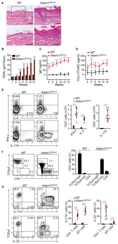
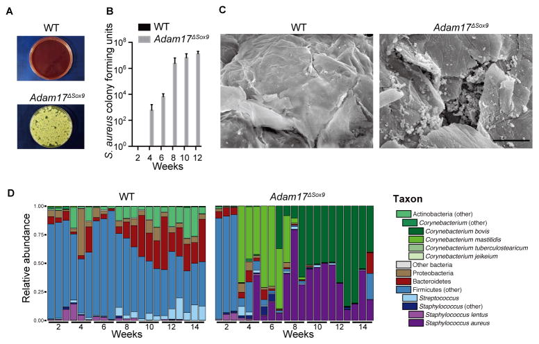
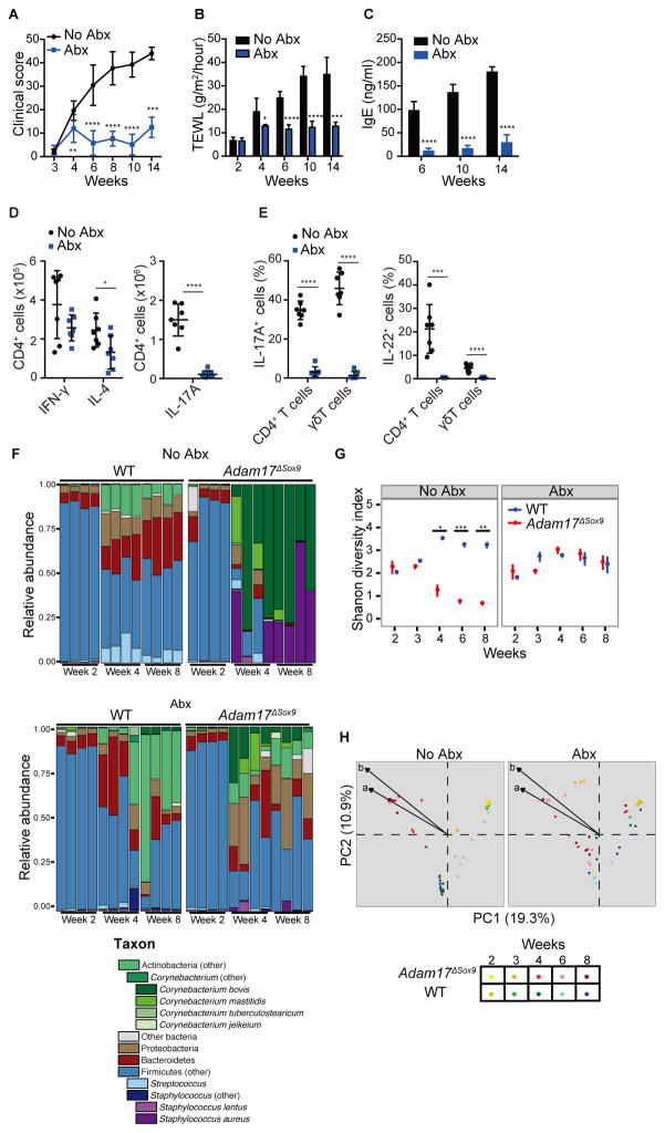
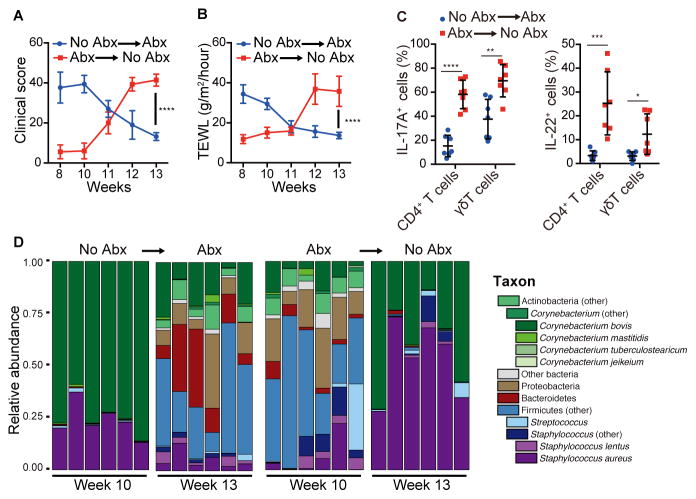
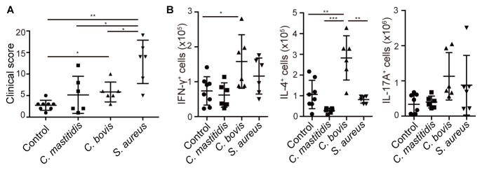
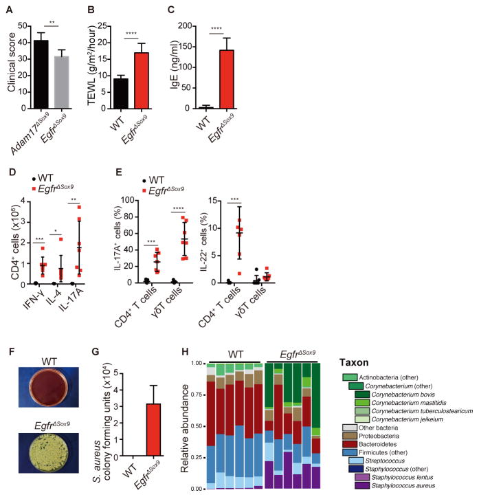
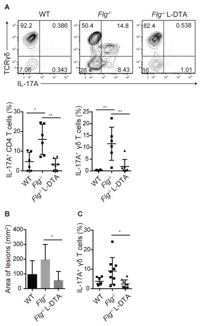
Staphylococcus aureus skin colonization is universal in atopic dermatitis and common in cancer patients treated with epidermal growth factor receptor inhibitors. However, the causal relationship of dysbiosis and eczema has yet to be clarified. Herein, we demonstrate that Adam17(fl/fl)Sox9-(Cre) mice, generated to model ADAM17-deficiency in human, developed eczematous dermatitis with naturally occurring dysbiosis, similar to that observed in atopic dermatitis. Corynebacterium mastitidis, S. aureus, and Corynebacterium bovis sequentially emerged during the onset of eczematous dermatitis, and antibiotics specific for these bacterial species almost completely reversed dysbiosis and eliminated skin inflammation. Whereas S. aureus prominently drove eczema formation, C. bovis induced robust T helper 2 cell responses. Langerhans cells were required for eliciting immune responses against S. aureus inoculation. These results characterize differential contributions of dysbiotic flora during eczema formation, and highlight the microbiota-host immunity axis as a possible target for future therapeutics in eczematous dermatitis.


Figures

Figure 1
Figure 1. Eczematous inflammation in ADAM17-deficient skin
(A) Histopathology of skin biopsy from WT and Adam17fl/flSox9-Cre (Adam17ΔSox9) mice. Scale bars, 50 μm. (B–D) TEWL, serum IgE and serum CCL17/TARC values in WT and Adam17ΔSox9 mice (N=8). (E) Flow cytometry analysis of IFN-γ, IL-4 and IL-17A production in CD4+ cells from lymph nodes, and (F, G) IL-17A and IL-22 production by CD3+ cells in epidermis of WT and Adam17ΔSox9 mice (N=8). TCRγδhigh cells represent dendritic epidermal T cells, and TCRγδmid cells represent γδ T cells that have infiltrated epidermis from the dermis. Data in B–G are shown as mean ± SD. *P<0.05, **P<0.01, ***P<0.001, ****P<0.0001 as determined by Student’s t test. See also Figures S1.
Figure 2
Figure 2. Staphylococcus aureus-associated dysbiosis in Adam17ΔSox9 mouse skin
(A, B) Quantification of S. aureus cultured from facial skin swab samples of WT and Adam17ΔSox9 mice (N=8, mean ± SD). (C) Scanning electron microscopy on skin surface of WT and Adam17ΔSox9 mice. Scale bars, 10 μm. (D) Microbiota of WT and Adam17ΔSox9 mice analyzed at indicated time points after birth. Relative abundance of order-genera that represented > 1% of total 16S rRNA gene sequences with additional speciation of Corynebacterium and Staphylococcus genus. Results represent two independent experiments (N=3). See also Figures S2.
Figure 3
Figure 3. Antibiotics prevent eczematous inflammation and conserves bacterial diversity
Adam17ΔSox9 mice were either treated or untreated with antibiotics cocktail (Abx) since weaning. See also Figure S3. (A–C) Clinical score, TEWL and serum IgE concentrations (N=7). A C shown as mean ± SD. *P<0.05, **P<0.01, ***P<0.001, ****P <0.0001 as determined by Student’s t test. (D) Flow cytometry analysis of IFN-γ, IL-4 and IL-17A production in CD4+ cells from lymph nodes, and (E) IL-17A and IL-22 production by CD3+ cells in epidermis of WT and Adam17ΔSox9 mice (N=7). (F) Microbiota of Abx-treated and untreated Adam17ΔSox9 and WT mice at indicated time points after birth. Relative abundance plots of order-genera that represented >1% of total 16S rRNA gene sequences with additional speciation of Corynebacterium and Staphylococcus genus of 4 representative mice for each time point. Results are representative of two experiments. (G) Shannon diversity index of Abx-treated and untreated Adam17ΔSox9 and WT mice from week 2 to 8 after birth. Each time point represents mean ± SEM. *P<0.05, **P<0.01, ***P<0.001 determined by Student’s t test and adjusted for multiple comparison by Benjamini and Hochberg correction. (H) Principal coordinates analysis (PCoA) of the microbial community structure (Theta similarity index) of Abx-treated and untreated Adam17ΔSox9 and WT mice. A shorter distance between points indicates higher similarity between the microbiome of samples represented by these points. Biplot arrows indicate the two most significant unique consensus taxonomies contributing to variation along axes 1 and 2 as determined by Spearman correlation. Length of biplot arrows reflects the contribution of that taxa to the top two axes: a, S. aureus (r=−0.73, P<1e-15); b, C. bovis (r=−0.77, P<1e-15). Spearman correlations and P-values refer to axis 1.
Figure 4
Figure 4. Antibiotics have therapeutic effect in Adam17ΔSox9 mice
Adam17ΔSox9 mice were untreated from 3~9 weeks after birth, and were put on antibiotics from 10~13 weeks, or vice versa. See also Figure S4. (A, B) Clinical score and TEWL in Adam17ΔSox9 mice during the crossover protocol (N=7). (C) IL-17A and IL-22 production by CD3+ cells in epidermis of Adam17ΔSox9 mice during the crossover protocol (N=7). (D) Relative abundance of order-genera that represented >1% of total 16S rRNA sequences with additional speciation of Corynebacterium and Staphylococcus genus before and after crossover. The data in A–C are shown as the mean ± SD. *P<0.05, **P<0.01, ***P<0.001, ****P<0.0001 determined by Student’s t test.
Figure 5
Figure 5. Contribution of S. aureus, C. bovis and C. mastitidis to eczematous inflammation
After withdrawal of antibiotic treatment form Adam17ΔSox9 mice that were pretreated with antibiotics, S. aureus, C. bovis and C. mastitidis strains were inoculated onto the mice. See also Figure S5. (A) Clinical scores of Adam17ΔSox9 mice after bacterial inoculation (N=6–8). (B) Flow cytometry analysis of IFN-γ, IL-4 and IL-17A production in CD4+ cells from lymph nodes of bacteria-inoculated Adam17ΔSox9 mice (N=6–8). The data are shown as the mean ± SD. *P<0.05, **P<0.01, ***P<0.001 determined by Student’s t test.
Figure 6
Figure 6. Ablation of EGFR-signaling induces dysbiosis and eczematous inflammation
(A) Clinical scores in Adam17ΔSox9 and EgfrΔSox9 mice (N=7). See also Figure S6. (B, C) TEWL and serum IgE values in WT and EgfrΔSox9 mice (N=7). (D) Flow cytometry analysis of IFN-γ, IL-4 and IL-17A production in CD4+ cells from lymph nodes, and (E) IL-17A and IL-22 production by CD3+ cells in epidermis of WT and EgfrΔSox9 mice (N=7). (F, G) Quantification of S. aureus cultured from facial skin swab samples of WT and EgfrΔSox9 mice (N=7). (H) Microbiota of 8-week-old WT and Egfr17ΔSox9 mice.
Figure 7
Figure 7. Langerhans cells mediate IL-17 response upon S. aureus inoculation
(A) Flow cytometry analysis of epidermis-infiltrating CD4 T cells and γδ T cells in topical inoculation of S. aureus on WT, Flg−/− and Flg−/− Langerin-DTA mice (N=6). (B, C) Area of inflamed lesions and epidermis-infiltrating γδ T cells of WT, Flg−/− and Flg−/− Langerin-DTA mice inoculated with S. aureus via occlusive dressing technique. The data are shown as the mean ± SD. *P<0.05, **P<0.01 determined by Student’s t test. See also Figure S7.

References

    1. Bath-Hextall FJ, Birnie AJ, Ravenscroft JC, Williams HC. Interventions to reduce Staphylococcus aureus in the management of atopic eczema: an updated Cochrane review. Br J Dermatol. 2010;163:12–26. - PubMed
    1. Blaydon DC, Biancheri P, Di WL, Plagnol V, Cabral RM, Brooke MA, van Heel DA, Ruschendorf F, Toynbee M, Walne A, et al. Inflammatory skin and bowel disease linked to ADAM17 deletion. N Engl J Med. 2011;365:1502–1508. - PubMed
    1. Blobel CP. ADAMs: key components in EGFR signalling and development. Nat Rev Mol Cell Biol. 2005;6:32–43. - PubMed
    1. Chavanas S, Bodemer C, Rochat A, Hamel-Teillac D, Ali M, Irvine AD, Bonafe JL, Wilkinson J, Taieb A, Barrandon Y, et al. Mutations in SPINK5, encoding a serine protease inhibitor, cause Netherton syndrome. Nat Genet. 2000;25:141–142. - PubMed
    1. Chey WD, Wong BC. American College of Gastroenterology guideline on the management of Helicobacter pylori infection. Am J Gastroenterol. 2007;102:1808–1825. - PubMed

Publication types

MeSH terms

Associated data