. 2015 Jun;123(3):140-8.
doi: 10.1111/eos.12184. Epub 2015 Apr 22.

Localization of cystic fibrosis transmembrane conductance regulator signaling complexes in human salivary gland striated duct cells

Affiliations

Localization of cystic fibrosis transmembrane conductance regulator signaling complexes in human salivary gland striated duct cells

Vina Z Zinn et al. Eur J Oral Sci. 2015 Jun.

Abstract

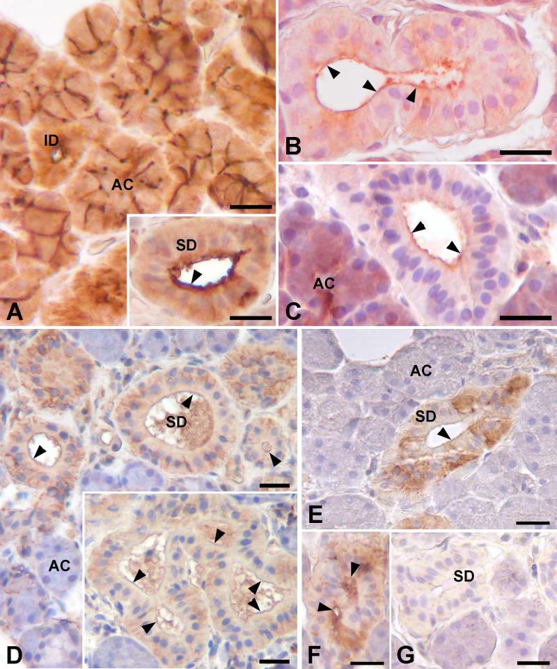
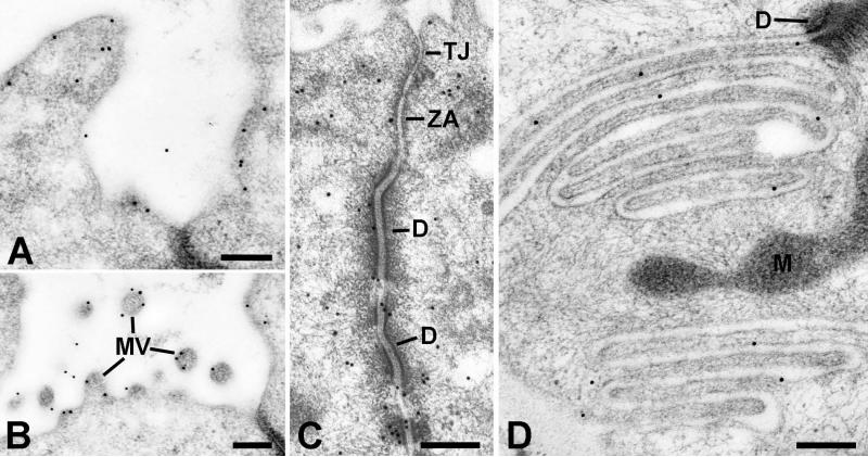
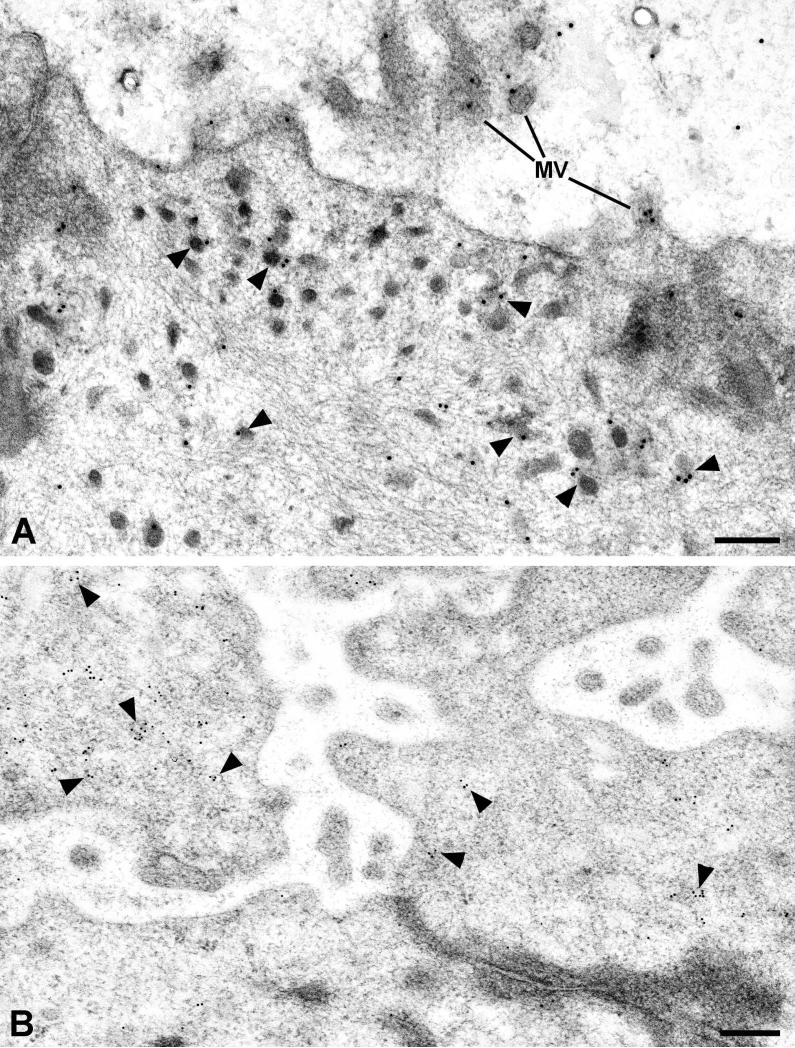
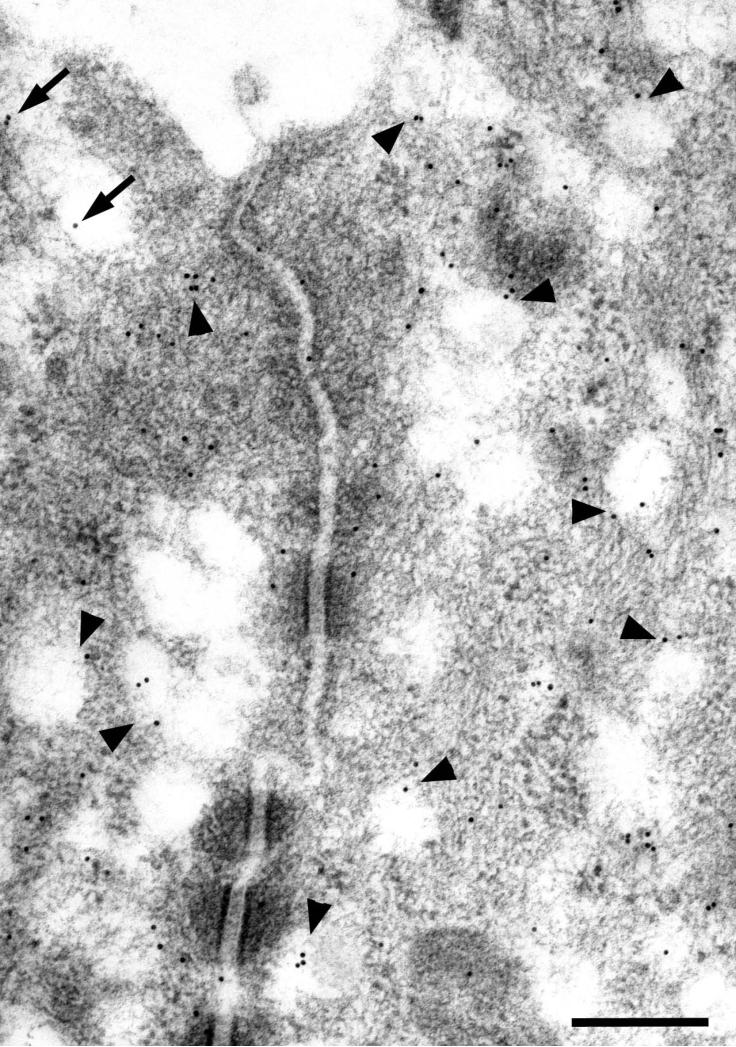
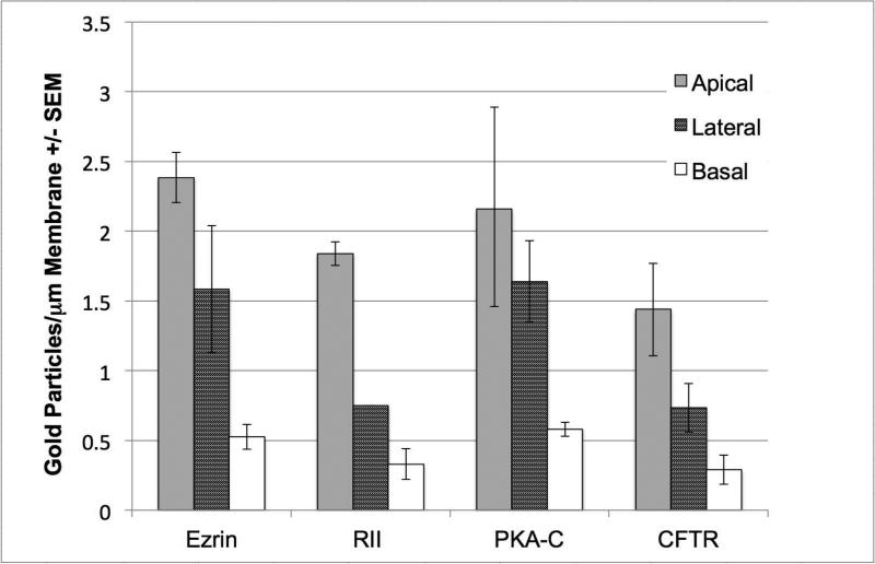
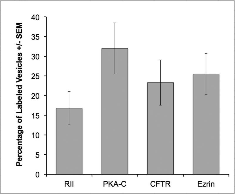
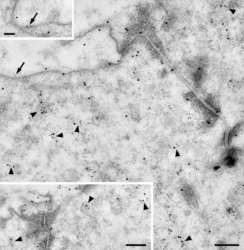
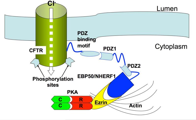
The cystic fibrosis transmembrane conductance regulator (CFTR) is a cyclic AMP-dependent protein kinase (PKA)-regulated Cl(-) channel, crucial for epithelial cell regulation of salt and water transport. Previous studies showed that ezrin, an actin binding and A-kinase anchoring protein (AKAP), facilitates association of PKA with CFTR. We used immunohistochemistry and immunogold transmission electron microscopy to localize CFTR, ezrin, and PKA type II regulatory (RII) and catalytic (C) subunits in striated duct cells of human parotid and submandibular glands. Immunohistochemistry localized the four proteins mainly to the apical membrane and the apical cytoplasm of striated duct cells. In acinar cells, ezrin localized to the luminal membrane, and PKA RII subunits were present in secretory granules, as previously described. Immunogold labeling showed that CFTR and PKA RII and C subunits were localized to the luminal membrane and associated with apical granules and vesicles of striated duct cells. Ezrin was present along the luminal membrane, on microvilli and along the junctional complexes between cells. Double labeling showed specific protein associations with apical granules and vesicles and along the luminal membrane. Ezrin, CFTR, and PKA RII and C subunits are co-localized in striated duct cells, suggesting the presence of signaling complexes that serve to regulate CFTR activity.

Keywords: immunohistochemistry; parotid gland; protein kinase A; submandibular gland; transmission electron microscopy.


Figures

Figure 1
Figure 1
Light microscopic localization of ezrin, PKA and CFTR in human salivary glands. A: SMG; ezrin reactivity is present around the lumen and along the intercellular canaliculi in secretory acini (AC), and around the lumen of intercalated duct (ID) cells. Inset: Parotid; ezrin is present along the luminal surface of striated duct cells (SD). B, C: In striated duct cells, RII subunits of PKA are present along the luminal membrane (arrowheads) and in the apical cytoplasm; in some cells reactivity also is present in the basal cytoplasm. In acinar cells, RII is present mainly in the apical region. B, SMG; C, parotid gland. D: SMG; C subunits of PKA are present along the luminal membrane (arrowheads) and in the basal cytoplasm of striated duct cells. E: SMG; CFTR is present along the luminal membrane (arrowheads) of striated duct cells, in the apical cytoplasm, and in the basal cytoplasm of some cells. No reactivity is present in the acini. F: Parotid; CFTR reactivity is present along the luminal membrane (arrowheads) and in the apical cytoplasm of striated duct cells. G: SMG; no labeling is seen in control sections incubated with non-immune IgG instead of primary antibody. Scale bars = 20 μm.
Figure 2
Figure 2
Electron microscopic localization of ezrin in striated duct cells. Gold particles indicating ezrin reactivity are associated with the luminal membrane (A) and microvilli (MV) (B), the lateral membranes especially in relation to intercellular junctions and associated cytoskeletal components (C), and the basal membrane infoldings (D). Tight junction (TJ); zonula adherens (ZA); desmosome (D); mitochondrion (M). Scale bars = 0.25 μm.
Figure 3
Figure 3
Electron microscopic localization of PKA in striated duct cells. A: RII subunits of PKA are present along the luminal membrane and microvilli (MV), and are associated with apical granules and vesicles (arrowheads). B: Catalytic (C) subunits of PKA are mainly associated with apical granules and vesicles. Scale bars = 0.25 μm.
Figure 4
Figure 4
CFTR is mainly associated with apical granules and vesicles of striated duct cells. The arrows indicate particles associated with the apical membrane. Scale bar = 0.25 μm.
Figure 5
Figure 5
Immunogold labeling of striated duct cell plasma membranes. Quantitative analysis showed that the labeling density of all four proteins was greatest for the apical membrane, and that a gradual decrease in density occurs from apical to lateral to basal membranes.
Figure 6
Figure 6
Percentage (± SEM) of apical granules/vesicles in striated duct cells labeled for RII, PKA-C, CFTR or ezrin.
Figure 7
Figure 7
Double labeling for RII (15 nm gold particles) and CFTR (10 nm gold particles) in parotid striated duct cells. RII is present along the luminal membrane and associated with apical granules and vesicles. CFTR is mainly associated with granules and vesicles; gold particles representing CFTR also are associated with the luminal membranes (arrows). Arrowheads indicate granules and vesicles labeled for both RII and CFTR. Scale bars = 0.25 μm; upper inset = 0.1 μm.
Figure 8
Figure 8
Schematic depicting possible CFTR-PKA-ezrin interaction at the apical membrane of striated duct cells allowing for rapid and efficient activation of the CFTR channel. PKA is anchored to ezrin via its RII subunits; ezrin in turn binds to EBP50/NHERF1, which has 2 PDZ domains that interact with the PDZ binding motif at the C-terminus of CFTR. Ezrin also binds to actin, which stabilizes the entire complex near the luminal membrane. Not shown are the heterotrimeric Gs protein (48) and other components of signaling pathways demonstrated (in other systems) or presumed to be involved with CFTR localization and activation (β-adrenergic receptor, P2Y2 receptor, adenylate cyclase, protein kinase C, etc.).

References

    1. WELSH MJ, BOAT TF, TSUI L-C, BEAUDET AL. Cystic Fibrosis. In: Seriven CR, Beaudet AL, Sly WS, Valle D, editors. The Metabolic Basis of Inherited Disease. McGraw Hill; New York, NY: 1995. pp. 3799–3876.
    1. FRIZZELL RA, HANRAHAN JW. Physiology of epithelial chloride and fluid secretion. Cold Spring Harb Perspect Med. 2012 doi: 10.1101/cshperspect.a009563. - PMC - PubMed
    1. LI C, NAREN AP. CFTR chloride channel in the apical compartments: spatiotemporal coupling to its interacting partners. Integr Biol (Camb) 2010;2:161–177. - PMC - PubMed
    1. PRANKE IM. SERMET-GAUDELUS I. Biosynthesis of cystic fibrosis transmembrane conductance regulator. Int J Biochem Cell Biol. 2014 http://dx.doi.org/10.1016/j.biocel.2014.03.020. - DOI - PubMed
    1. CANT N, POLLOCK N, FORD RC. CFTR structure and cystic fibrosis. Int J Biochem Cell Biol. 2014 http://dx.doi.org/10.1016/j.biocel.2014.02.004. - DOI - PubMed

Publication types

MeSH terms

Substances