. 2015 May 29;290(22):14235-44.
doi: 10.1074/jbc.M114.621615. Epub 2015 Apr 22.

EPHB4 Protein Expression in Vascular Smooth Muscle Cells Regulates Their Contractility, and EPHB4 Deletion Leads to Hypotension in Mice

Affiliations

EPHB4 Protein Expression in Vascular Smooth Muscle Cells Regulates Their Contractility, and EPHB4 Deletion Leads to Hypotension in Mice

Yujia Wang et al. J Biol Chem. .

Abstract

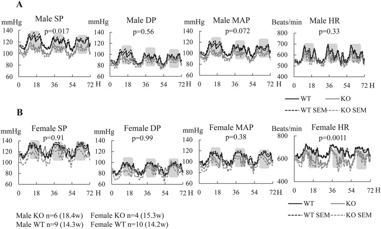
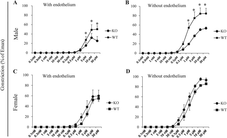
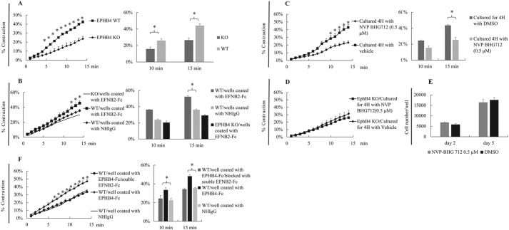
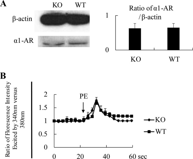
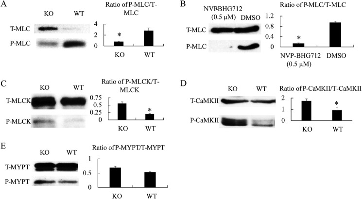
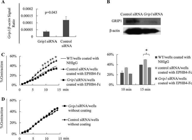
EPH kinases are the largest family of receptor tyrosine kinases, and their ligands, ephrins (EFNs), are also cell surface molecules. This work presents evidence that EPHB4 on vascular smooth muscle cells (VSMCs) is involved in blood pressure regulation. We generated gene KO mice with smooth muscle cell-specific deletion of EPHB4. Male KO mice, but not female KO mice, were hypotensive. VSMCs from male KO mice showed reduced contractility when compared with their WT counterparts. Signaling both from EFNBs to EPHB4 (forward signaling) and from EPHB4 to EFNB2 (reverse signaling) modulated VSMC contractility. At the molecular level, the absence of EPHB4 in VSMCs resulted in compromised signaling from Ca(2+)/calmodulin-dependent protein kinase II (CaMKII) to myosin light chain kinase (MLCK) to myosin light chain, the last of which controls the contraction force of motor molecule myosin. Near the cell membrane, an adaptor protein GRIP1, which can associate with EFNB2, was found to be essential in mediating EPHB4-to-EFNB reverse signaling, which regulated VSMC contractility, based on siRNA gene knockdown studies. Our research indicates that EPHB4 plays an essential role in regulating small artery contractility and blood pressure.

Keywords: EPHB4 kinases; GRIP1; cell biology; gene knockout; hormone; hypertension; hypotension; sex hormones; vascular smooth muscle cells.


Figures

FIGURE 1.
FIGURE 1.
Generation of mice with SMC-specific Ephb4 null mutation. A, scheme of Ephb4f/f mouse generation. Bold lines represent the left and right arms of genomic sequences used in gene targeting. LoxP and FRT sites are represented by empty and gray small arrowheads, respectively. The hatched box represents a genomic region from which probes were produced for Southern blotting analysis. B, Southern blotting analysis of tail DNA of floxed Ephb4 mice. Tail DNA was digested with EcoRV/BclI or BamHI/EcoRI, and then hybridized with the 5′ probe or 3′ probe, respectively. For the 5′ probe (upper panel), the 11.4-kb band was derived from the WT allele, and the 9.1-kb band was derived from the mutated allele. For the 3′ probe (lower panel), the 12.6-kb band was derived from the WT allele, and the 7.0-kb band was derived from the mutated allele. C, Ephb4 mRNA deletion in mesenteric arteries of Ephb4 KO mice. RNA was extracted from mesenteric arteries and spleens from WT and Ephb4 KO mice and analyzed by RT-qPCR for Ephb4 mRNA levels. β-Actin mRNA levels were used as an internal control. Samples in RT-qPCR were in triplicate, and means ± S.E. of Ephb4 signal/β-actin signal ratios are shown. The experiment was conducted twice; a representative one is shown. D, EPHB4 protein deletion in EPHB4 KO VSMCs according to immunoblotting. VSMCs from Ephb4 KO and WT mice were cultured for 4 days and then harvested. Cell lysates were analyzed for EPHB4 protein expression by immunoblotting. Spleens from KO and WT mice were used as controls for tissue specificity. The experiment was conducted twice; a representative one is shown.
FIGURE 2.
FIGURE 2.
Male EPHB4 KO mice were hypotensive. BP and HR were measured for 72 h by radiotelemetry, starting at least 7 days after radio transmitter implantation. The number of mice per group and their mean age at the time of BP measurement are indicated. Means ± S.E. of hourly BP and HR during the 72-h period are presented. Shaded areas represent dark periods. MAP, mean arterial pressure. The statistical significance of differences between BP and HR of the experimental groups was evaluated by analysis of variance, and p values are indicated. A, BP and HR of males. B, BP and HR of females.
FIGURE 3.
FIGURE 3.
Reduced contractility of mesenteric arteries from male EPHB4 KO mice. A–D, segments of the third-order branch of the mesenteric artery with (A and C) or without (B and D) endothelium were stimulated with PE. A single cumulative concentration-response curve to PE (1 nm to 100 μm) was obtained. Maximal tension (Emax) was determined by challenging the vessels with physiological saline containing 127 mm KCl. Vessel contractility is expressed as the percentage of Emax. Data from two mice (with three arterial segments tested for each mouse, i.e. six determinants per group) were pooled, and means ± S.E. are reported. Contractility differences were analyzed by paired Student's t tests, and p values are indicated. *, p < 0.05. A, contractility of mesenteric arteries with endothelium from male KO and WT mice. B, contractility of mesenteric arteries without endothelium from male KO and WT mice. C, contractility of mesenteric arteries with endothelium from female KO and WT mice. D, contractility of mesenteric arteries without endothelium from female KO and WT mice.
FIGURE 4.
FIGURE 4.
VSMCs stimulated by both EPHB4 forward and EPHB4 reverse signaling show increased contractility. VSMCs from male WT and KO mice were cultured for 4 days. In some experiments, the VSMCs were cultured in wells coated with EFNB2-Fc (2 μg/ml for coating), EPHB4-Fc (2 μg/ml for coating), or NHIgG (2 μg/ml for coating, as control for EPHB4-Fc), as indicated. VSMCs were stimulated with 20 μm PE at 37 °C and imaged every min for 15 min. All experiments were conducted 3 times independently. Means ± S.E. of percentage of contraction of 15–30 cells in a representative experiment are shown. Data at 10 and 15 min of 3 independent experiments are summarized and expressed as bar graphs (means ± S.E.) on the left. The data were analyzed by paired Student's t test (*, p < 0.05). WT, WT VSMC; KO, KO VSMC. A, decreased contractility of VSMCs from male EPHB4 KO mice. VSMCs were cultured in plain wells. B, forward signaling through EPHB4 increases WT VSMC contractility. WT VSMCs were cultured in wells coated with EFNB2-Fc. KO VSMCs in coated wells were used as negative controls. WT VSMCs cultured in wells coated with NHIgG were used as additional controls, and their mean contractility is presented as a thin line without S.E. to facilitate viewing. C, EPHB4 inhibitor NVP-BHG712 suppresses WT VSMC contractility. WT VSMCs were cultured in plain wells in the presence of NVP-BHG712 (0.5 μm) or vehicle for the last 4 h of culture. Their contractility after PE stimulation was measured. D, EPHB4 inhibitor NVP-BHG712 does not affect KO VSMC contractility. KO VSMCs were cultured in plain wells in the presence of NVP-BHG712 (0.5 μm) or vehicle for the last 4 h of culture. Their contractility after PE stimulation was measured. E, VSMCs cultured in the presence of EPHB4 inhibitor NVP-BHG712 show normal proliferation. VSMCs from WT male mice were cultured for 2–5 days in the presence of NVP-BHG712 at 0.5 μm as indicated. Cell number per well of 24-well plate at days 2 and 5 was enumerated. Means ± S.E. of cell counting of three wells are shown. The experiment was conducted twice; data from a representative experiment are shown. No significant difference was found between NVP-BHG712- and vehicle-treated cells (Student's t test). F, reverse signaling triggered by EPHB4 increases WT VSMC contractility. WT VSMCs were cultured in wells coated with EPHB4-Fc. In one of the groups, soluble EFNB2-Fc (2 μg/ml) was added to culture to block interaction between solid-phase EPHB4-Fc and cell surface EFNB2. WT VSMCs cultured in wells coated with NHIgG were used as an additional control, and their mean contractility is presented as a thin line without S.E. to facilitate viewing.
FIGURE 5.
FIGURE 5.
AR and Ca2+ flux in KO VSMCs. A, normal α1-AR expression in EPHB4 KO VSMCs according to immunoblotting. VSMCs from male Ephb4 KO and WT mice were cultured for 4 days and then harvested. Cell lysates were analyzed for α1-AR expression by immunoblotting. β-Actin levels were used as internal controls. A representative immunoblot is shown. Densitometry data from 2 independent experiments were pooled and presented as a bar graph on the right with means ± S.E. indicated. No statistically significant differences between KO and WT VSMCs were found according to Student's t test. B, normal Ca2+ flux in VSMCs from EPHB4 KO mice. VSMCs from male Ephb4 KO or WT mice were cultured for 4 days and loaded with Fura2. They were then placed in Hanks' balanced salt solution without Ca2+ at 37 °C and stimulated with PE (20 μm). The arrow indicates the time point at which PE was added. The ratio of emissions at 510 nm triggered by 340 nm versus 380 nm excitation in each cell was registered every 3 s for 1 min. The experiments were conducted 3 times. Means ± S.E. of the ratio of more than 15 randomly selected VSMCs in a representative experiment are illustrated. No statistically significant differences between the KO and WT groups were found according to Student's t test.
FIGURE 6.
FIGURE 6.
MLC, MLCK, CaMKII, and MYPT phosphorylation of VSMC from WT and KO mice. VSMCs from male KO and WT mice were cultured for 4 days and then stimulated with 20 μm PE for 3 s and immediately lysed. Total (T-) and phosphorylated (P-) MLC, MLCK, and CaMKII were analyzed by immunoblotting. Three independent experiments were conducted. Immunoblotting images from representative experiments are illustrated. The signal ratios of phosphorylated versus total MLC, MLCK, CaMKII, and MYPT were quantified by densitometry. Densitometry data from the 3 independent experiments were pooled and are presented as bar graphs (means ± S.E. of relative intensity) in the panels on the left. Paired Student's t tests were employed for statistical analysis. * indicates p < 0.05. A, decreased MLC phosphorylation in VSMC from EPHB4 KO mice. B, decreased MLC phosphorylation in VSMCs after EPHB4 inhibitor treatment. WT VSMCs were cultured in the presence of NVP-BHG712 (0.5 μm) for the last 4 h of culture. The controls were treated with vehicle (dimethyl sulfoxide (DMSO)) at the same dilution and duration. C, increased MLCK phosphorylation in VSMCs from EPHB4 KO mice. D, increased CaMKII phosphorylation in VSMCs from EPHB4 KO mice. E, MYPT phosphorylation remains unchanged in VSMCs from EPHB4 KO mice.
FIGURE 7.
FIGURE 7.
GRIP1 mediates EPHB4 reverse signaling in controlling VSMC contractility. A, effective Grip1 mRNA knockdown by siRNA. Cultured WT VSMCs were transfected with a mixture of three pairs of Grip1 siRNAs or control siRNA, as indicated. After a 24-h culture, the cells were harvested, and the mRNA expression was determined by RT-qPCR, samples being in duplicate. The experiments were conducted twice, and the data (four determinants for each group) were pooled and expressed as means ± S.E. of the ratios of the target gene signal versus the β-actin signal. The data were analyzed by Student's t test. *, p < 0.05. B, effective GRIP1 protein knockdown by siRNA. The cells as described in panel A were analyzed by immunoblotting for GRIP1 protein expression. β-Actin was used as loading control. Experiments were conducted 3 times, and representative ones are shown. C, GRIP1 knockdown by siRNAs partially reverses the enhancing effect of solid-phase EPHB4-Fc in VSMC contractility. VSMCs from WT males were cultured in wells coated with recombinant EPHB4-Fc (2 μg/ml during coating). After 2 days, they were transfected with siRNAs targeting Grip1 mRNA, or with control siRNA. On day 4 of culture, they were stimulated with PE (20 μm), and their percentage of contraction was recorded. Means ± S.E. of percentages are reported. The thin lines represent the mean percentage of contraction of VSMCs cultured in wells coated with NHIgG (2 μg/ml) without siRNA transfection (for a better visual effect, the S.E. of each time point in this control is omitted). All experiments were conducted 3 times independently. Means ± S.E. of percentage of contraction of 15–30 cells of a representative experiment are shown. The data at 10 and 15 min of 3 independent experiments are summarized and expressed as bar graphs (means ± S.E.) on the left. The data were analyzed by paired Student's t test (*, p < 0.05). D, GRIP1 knockdown in VSMCs without EPHB4 reverse signaling does not affect VSMC contractility. WT VSMCs were cultured in plain wells and transfected with Grip1 or control siRNA as described for panel C. Their contractility upon PE stimulation was measured. No significant differences were observed according to Student's t test.

References

    1. Eph Nomenclature Committee (1997) Unified nomenclature for Eph family receptors and their ligands, the ephrins. Cell 90, 403–404 - PubMed
    1. Pasquale E. B. (2008) Eph-ephrin bidirectional signaling in physiology and disease. Cell 133, 38–52 - PubMed
    1. Wu J., Luo H. (2005) Recent advances on T-cell regulation by receptor tyrosine kinases. Curr. Opin. Hematol. 12, 292–297 - PubMed
    1. Flanagan J. G., Vanderhaeghen P. (1998) The ephrins and Eph receptors in neural development. Annu. Rev. Neurosci. 21, 309–345 - PubMed
    1. Luo H., Charpentier T., Wang X., Qi S., Han B., Wu T., Terra R., Lamarre A., Wu J. (2011) Efnb1 and Efnb2 proteins regulate thymocyte development, peripheral T cell differentiation, and antiviral immune responses and are essential for interleukin-6 (IL-6) signaling. J. Biol. Chem. 286, 41135–41152 - PMC - PubMed

Publication types