. 2015 Jun 12;290(24):15092-101.
doi: 10.1074/jbc.M115.653485. Epub 2015 Apr 21.

Olfactomedin-1 Has a V-shaped Disulfide-linked Tetrameric Structure

Affiliations

Olfactomedin-1 Has a V-shaped Disulfide-linked Tetrameric Structure

Matti F Pronker et al. J Biol Chem. .

Abstract

Olfactomedin-1 (Olfm1; also known as noelin and pancortin) is a member of the olfactomedin domain-containing superfamily and a highly expressed neuronal glycoprotein important for nervous system development. It binds a number of secreted proteins and cell surface-bound receptors to induce cell signaling processes. Using a combined approach of x-ray crystallography, solution scattering, analytical ultracentrifugation, and electron microscopy we determined that full-length Olfm1 forms disulfide-linked tetramers with a distinctive V-shaped architecture. The base of the "V" is formed by two disulfide-linked dimeric N-terminal domains. Each of the two V legs consists of a parallel dimeric disulfide-linked coiled coil with a C-terminal β-propeller dimer at the tips. This agrees with our crystal structure of a C-terminal coiled-coil segment and β-propeller combination (Olfm1(coil-Olf)) that reveals a disulfide-linked dimeric arrangement with the β-propeller top faces in an outward exposed orientation. Similar to its family member myocilin, Olfm1 is stabilized by calcium. The dimer-of-dimers architecture suggests a role for Olfm1 in clustering receptors to regulate signaling and sheds light on the conformation of several other olfactomedin domain family members.

Keywords: analytical ultracentrifugation; cell signaling; coiled coil; development; disulfide; electron tomography; neurobiology; olfactomedin-1 (Olfm1); small angle x-ray scattering (SAXS); x-ray crystallography.


Figures

FIGURE 1.
FIGURE 1.
Olfm1 forms disulfide-linked tetramers. A, isoforms and domains of Olfm1. In this study isoform 1 was used. Cysteines are shown as vertical black lines. SP, signal peptide. B, non-reducing SDS-PAGE (left, Coomassie-stained purified protein; right, Western blotted expression medium) shows a shift to >250 kDa, whereas reducing SDS-PAGE shows Olfm1 running at the expected mass of 64 kDa for a (fully glycosylated) monomer (M). T, tetramer; βME, β-mercaptoethanol; lane M, molecular mass markers. C, SEC-MALS shows a mass of ∼260 kDa for the Olfm1 peak, which elutes before conalbumin (Conalb.) (78 kDa). The plotted UV signal was recorded at 280 nm. AU, absorbance units. D, AUC sedimentation velocity experiments give a mass of 242 kDa, a sedimentation coefficient of 7.67 S, and a frictional ratio of 1.98 (root mean square deviation, 0.005). E, SAXS log I(s) versus s plot of Olfm1 at 3.55 mg/ml. F, Guinier plot from SAXS curve agrees with a tetramer (I0 = 249, Rg = 8.5 nm).
FIGURE 2.
FIGURE 2.
A, structure of the Olfm1coil-Olf dimer. The inter- and intraprotein disulfides (shown as spheres) link the two monomers together and lock the β-propellers to the coiled coil, respectively. B, close-up view of the coiled coil and disulfides of the Olfm1coil-Olf dimer showing the hydrophobic side chains in the coiled coil as sticks. The 2FoFc electron density map was plotted at 1.2σ. C, view down the top face of a single β-propeller shows five blades, which are numbered accordingly. D, the structure of the olfactomedin domain of gliomedin (Gldn; red) is very similar to the olfactomedin domain of Olfm1 (teal; root mean square deviation of 1.3 Å over 226 aligned Cα atoms).
FIGURE 3.
FIGURE 3.
Analysis of the hydrophobic heptad repeat pattern in the coiled-coil domain of Olfm1. The full amino acid sequence of mouse Olfm1 is shown. Hydrophobic heptad repeat residues have been indicated based on the register of coiled-coil residues 210–225, which were observed in the crystal structure. Also indicated are possible disturbances from an ideal coiled coil such as frameshifts, hydrophilic residues on heptad repeat positions, and prolines in the coiled coil.
FIGURE 4.
FIGURE 4.
A, Olfm1coil-Olf dimer with modeled full glycans (red) and longer coiled coils shows that all glycans are localized to the side and bottom face of the β-propeller, whereas the top face is accessible. B, conservation plot indicates that the top face of the β-propeller has a higher degree of sequence conservation than the sides, suggesting that this interface might be important for ligand binding. Conservation scores were calculated with an alignment of 35 vertebrate Olfm1 orthologs (supplemental Fig. 1) using ConSurf (42).
FIGURE 5.
FIGURE 5.
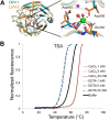
A, comparison of the calcium-binding olfactomedin domain of latrophilin3 (Lphn3; orange) and Olfm1 (teal). It is likely that Olfm1 can also accommodate a calcium ion at the center of the β-propeller. The latrophilin3-coordinated calcium ion (green) and neighboring sodium ion (purple) are shown. B, TSA shows a pronounced stabilizing effect of Olfm1 by calcium, shifting the melting temperature 8 °C higher, whereas EDTA destabilizes Olfm1 (a shift to 10.5 °C lower). Buffer indicates TSA buffer without calcium or EDTA added.
FIGURE 6.
FIGURE 6.
The full-length Olfm1 tetramer has a V-shaped architecture. A, the Kratky plot shows no signs of large unstructured regions (37). B, the SAXS pair distance distribution function P(r) indicates a dumbbell-like shape. C, ab initio modeling by DAMMIF with imposed 2-fold rotational symmetry reveals a V-shaped architecture. A single (non-averaged) DAMMIF model is shown that is representative for the average structure generated from 50 models without applying 2-fold symmetry. D, fit from the P(r) and the DAMMIF ab initio modeling (χ2 = 4.313). E, ET shows V shapes with varying angles between the legs. The dimensions of the coiled coil and the double β-propeller from the crystal structure fit those of the ET densities. Top panels, central slice through the tomograms showing the V shapes. Middle panels, fit of two double β-propellers with modeled coiled-coil domains in the ET density. Bottom panels, same as middle panels but without the ET densities. Scale bar, 10 nm. F, model of the full-length Olfm1 tetramer. Each monomer is represented by a different color. In the NTT domains, one of the interprotein disulfides necessary for tetramerization is indicated.
FIGURE 7.
FIGURE 7.
Sequence alignment of Olfm1 with the closely related (mouse) paralogs Olfm2, Olfm3, Olfm4, and myocilin. Domain boundaries are indicated with the same color scheme as in Fig. 1A. Conserved cysteines are indicated by arrowheads. Residues are colored according to the percentage of sequence identity (blue means conserved; white means variable). ER, endoplasmic reticulum.

References

    1. Snyder D. A., Rivers A. M., Yokoe H., Menco B. P., Anholt R. R. (1991) Olfactomedin: purification, characterization, and localization of a novel olfactory glycoprotein. Biochemistry. 30, 9143–9153 - PubMed
    1. Moreno T. A., Bronner-Fraser M. (2001) The secreted glycoprotein Noelin-1 promotes neurogenesis in Xenopus. Dev. Biol. 240, 340–360 - PubMed
    1. Moreno T. A., Bronner-Fraser M. (2005) Noelins modulate the timing of neuronal differentiation during development. Dev. Biol. 288, 434–447 - PubMed
    1. Rice H. C., Townsend M., Bai J., Suth S., Cavanaugh W., Selkoe D. J., Young-Pearse T. L. (2012) Pancortins interact with amyloid precursor protein and modulate cortical cell migration. Development 139, 3986–3996 - PMC - PubMed
    1. Barembaum M., Moreno T. A., LaBonne C., Sechrist J., Bronner-Fraser M. (2000) Noelin-1 is a secreted glycoprotein involved in generation of the neural crest. Nat. Cell Biol. 2, 219–225 - PubMed

Publication types

LinkOut - more resources