. 2015 Apr 23:6:6912.
doi: 10.1038/ncomms7912.

Superlattices assembled through shape-induced directional binding

Affiliations

Superlattices assembled through shape-induced directional binding

Fang Lu et al. Nat Commun. .

Abstract

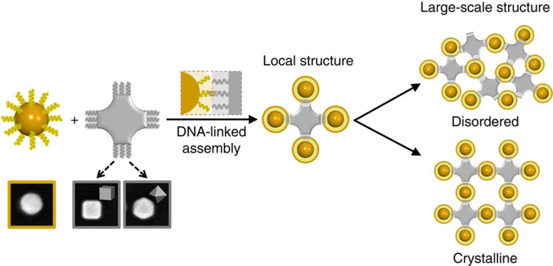
Organization of spherical particles into lattices is typically driven by packing considerations. Although the addition of directional binding can significantly broaden structural diversity, nanoscale implementation remains challenging. Here we investigate the assembly of clusters and lattices in which anisotropic polyhedral blocks coordinate isotropic spherical nanoparticles via shape-induced directional interactions facilitated by DNA recognition. We show that these polyhedral blocks--cubes and octahedrons--when mixed with spheres, promote the assembly of clusters with architecture determined by polyhedron symmetry. Moreover, three-dimensional binary superlattices are formed when DNA shells accommodate the shape disparity between nanoparticle interfaces. The crystallographic symmetry of assembled lattices is determined by the spatial symmetry of the block's facets, while structural order depends on DNA-tuned interactions and particle size ratio. The presented lattice assembly strategy, exploiting shape for defining the global structure and DNA-mediation locally, opens novel possibilities for by-design fabrication of binary lattices.


Figures

Figure 1
Figure 1. Schematic of shape-induced directional bonds for DNA-linked nanoparticle assembly.
SNPs (yellow unit) are isotropic objects with high symmetry. ANPs (grey unit) have low-order symmetry; for example, CB or OC as shown in electron micrographs (inner frame length of images is 100 nm). Regulated interactions between particles are induced by functionalizing SNPs and ANPs with complementary DNA strands. The directional bonding of ANPs coordinates SNPs in a local structure (clusters). Our study examines the conditions under which the shape of ANPs can be translated into well-established local structure, and factors affecting the formation of either disordered or ordered large-scale 3D assembly.
Figure 2
Figure 2. Cube-encoded assemblies of clusters with spherical nanoparticles.
(a; Top) SEM images of (from left to right) CBs (46 nm in edge length) and SNPs (46 nm in diameter); (bottom) schematic of the DNA functionalization and assembly of CB and SNP. (b) Comparison of (top) TEM images with (middle) 3D reconstruction models and (bottom) the projections of the modelled structures at a few selected tilt angles to reveal the 3D structure of the assembled clusters (from left to right: –72.1°, –52.1°, –38.5° and –18.1°). The projections of the modelled clusters agree with the recorded TEM images. (c; Top) Schematic and (bottom) SEM images illustrate the shape-induced directional bonding of CB in a cluster-dimension-extending history (from left to right). The scale bars, 50 nm.
Figure 3
Figure 3. Cube–sphere nanoparticle assemblies of superlattices.
(a) Schematic of a 46-nm SNP/46-nm CB pair linked by DNA system 18S. (b) SAXS data with experimental (blue) and modelled (red) structure factors, S(q), (c) scattering image and (d) the corresponding structure schematic for 46-nm SNP/46-nm CB assembly system, which crystallizes into a NaCl-type lattice with a space group symmetry of formula image
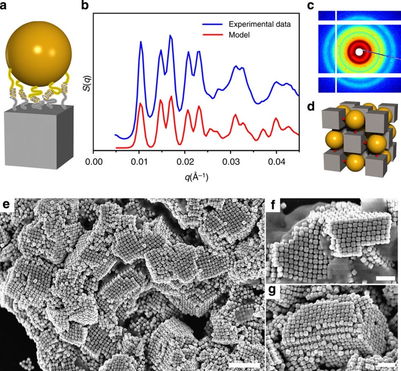
. (e) Low-magnification SEM image of SNP/CB-assembled crystals, where square-lattice ordering can be observed from the fragments, even though drying effects cause cracks in the crystals (scale bar, 500 nm). (f,g) High-magnification images of superlattice, demonstrating the alternate packing of SNPs and CBs in the 3D square lattice (scale bar, 200 nm).
Figure 4
Figure 4. Effects of DNA rigidity and sphere size on the structure of cube-sphere binary assembly.
(a) Schematic of a 46-nm SNP/46-nm CB pair linked by rigid DNA system 18R. (b) SAXS extracted structure factors S(q) and (inset) DNA-NP schematic (18R). (c,d) SEM image of 46-nm SNP/46-nm CB assemblies for rigid DNA system 18R. (e) Schematic of a 27-nm SNP/46-nm CB pair linked by flexible DNA system 18S. (f) SAXS data and (inset) DNA-NP schematic (18S). (g,h) Corresponding SEM. (i) Schematic of a 38-nm SNP/46-nm CB pair linked by flexible DNA system 18S. (j) SAXS data with experimental (blue) and modelled (red) structure factors, S(q), and (inset) scattering image, which crystallizes into a NaCl-type lattice. (k,l) Corresponding SEM images. Scale bars, 100 nm.
Figure 5
Figure 5. Synergetic effects of DNA shell and particle size mismatch on the structure of cube–sphere binary assembly.
(a) Low- and (b,c) high-magnification SEM images of binary superlattices formed by 38-nm SNPs/46-nm CBs with 30S DNA (left scale bar, 500 nm; right scale bar, 200 nm). (d) State diagram for all the studied binary systems of cubes, spheres and DNA designs. Leading parameters are the DNA length and flexibility, and the SNP/CB size ratio (CB is 46 nm in edge length), as discussed in the text. (e) Comparison between experimental (from SAXS data) and calculated (see Supplementary Note 4) values of the nearest-neighbour interparticle surface-to-surface distances, Dss, for 46-nm SNP/46-nm CB binary assembly (error bars correspond to one s.d. for repeated experiments). The calculated values account for the sum of DNA shell thicknesses on the cube and sphere surfaces, TDNA shell_cube+TDNA shell_sphere. (f) A normalized attractive potential energy as a function of the SNP displacement, ddis, from the CB axis that crosses the centre of the square face for SNPs with diameters of 46 nm (red line D1), 38 nm (orange line D2) and 27 nm (black line D3), respectively, and schematics of SNP/CB pair for calculations (inset, left for D1 and right for D3). (g) Assembly of similarly sized and dissimilar CB and SNP. The mechanism for order propagation from the cluster formation of similar-sized cubes and spheres (top), and disorder (bottom) is illustrated, see the text for details.
Figure 6
Figure 6. Octahedron-sphere nanoparticle assemblies.
(a; Top) SEM images of (from left to right) OCs (46 nm in edge length) and SNPs (46 nm in diameter); (bottom) schematic of DNA functionalization and assembly of OC and SNP. (b; Top) Schematic and (bottom) SEM images illustrating the directing role of octahedral blocks in the formation of clusters, and their merging into larger-scale structures (from left to right). Scale bars, 50 nm. (c) SAXS image and (d) data with experimental (blue) and modelled (red) structure factors, S(q). (e) The corresponding structure of formed CsCl-type superlattice with space group formula image 
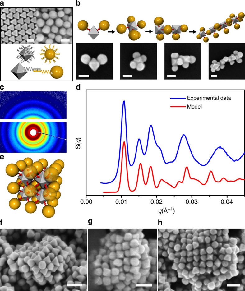
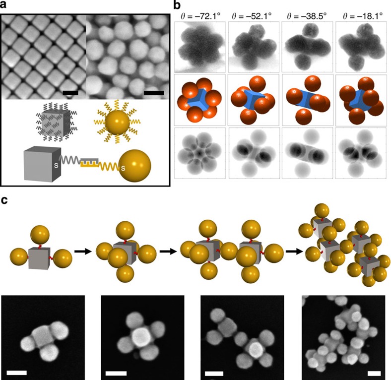
symmetry (left bottom) for 46-nm SNP/46-nm OC assemblies. (fh) SEM images of SNP/OC-assembled superlattice fragments (scale bar, 100 nm).

References

    1. Kuzyk A. et al.. DNA-based self-assembly of chiral plasmonic nanostructures with tailored optical response. Nature 483, 311–314 (2012). - PubMed
    1. Henry E. et al.. Crystallization of fluorescent quantum dots within a three-dimensional bio-organic template of actin filaments and lipid membranes. Nano. Lett. 11, 5443–5448 (2011). - PubMed
    1. Zhang Y. G., Lu F., Yager K. G., van der Lelie D. & Gang O. A general strategy for the DNA-mediated self-assembly of functional nanoparticles into heterogeneous systems. Nat. Nanotechnol. 8, 865–872 (2013). - PubMed
    1. Nykypanchuk D., Maye M. M., van der Lelie D. & Gang O. DNA-guided crystallization of colloidal nanoparticles. Nature 451, 549–552 (2008). - PubMed
    1. Macfarlane R. J. et al.. Nanoparticle superlattice engineering with DNA. Science 334, 204–208 (2011). - PubMed

Publication types